Review
. 2022 Aug;38(8):953-965.
doi: 10.1007/s12264-022-00845-6. Epub 2022 Mar 29.

Astrocytes in Post-traumatic Stress Disorder

Affiliations
Review

Astrocytes in Post-traumatic Stress Disorder

Baoman Li et al. Neurosci Bull. 2022 Aug.

Abstract

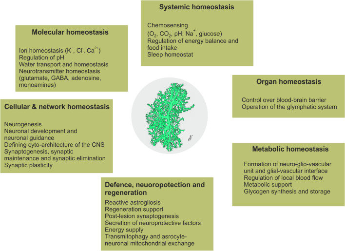
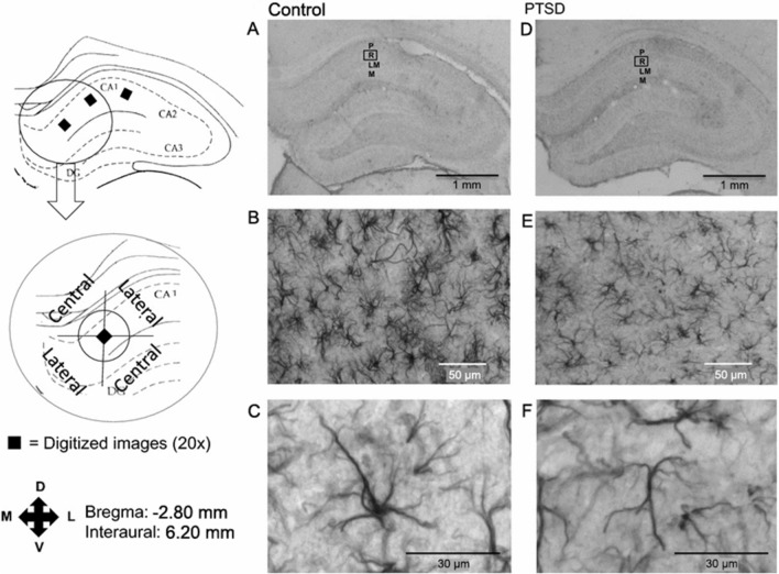
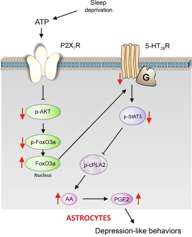
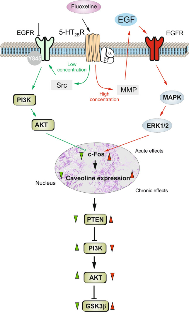
Although posttraumatic stress disorder (PTSD) is on the rise, traumatic events and their consequences are often hidden or minimized by patients for reasons linked to PTSD itself. Traumatic experiences can be broadly classified into mental stress (MS) and traumatic brain injury (TBI), but the cellular mechanisms of MS- or TBI-induced PTSD remain unknown. Recent evidence has shown that the morphological remodeling of astrocytes accompanies and arguably contributes to fearful memories and stress-related disorders. In this review, we summarize the roles of astrocytes in the pathogenesis of MS-PTSD and TBI-PTSD. Astrocytes synthesize and secrete neurotrophic, pro- and anti-inflammatory factors and regulate the microenvironment of the nervous tissue through metabolic pathways, ionostatic control, and homeostatic clearance of neurotransmitters. Stress or trauma-associated impairment of these vital astrocytic functions contribute to the pathophysiological evolution of PTSD and may present therapeutic targets.

Keywords: Astrocytes; Neurotrophic factors; Serotonin; Traumatic brain injury; Traumatic events.


Conflict of interest statement

The authors declare no competing interests.

Figures

Fig. 1
Fig. 1
Functions of astrocytes.
Fig. 2
Fig. 2
Digitized images of the hippocampus after GFAP immunohistochemistry showing the CA1 region. AC Control; DF PTSD. A, D Digitized images at 1×; B, E digitized images at 20×; C, F digitized images at 40×. Square areas denote regions of interest at 20×. P, stratum pyramidale; R, stratum radiatum; LM, stratum lacunosum moleculare; M, stratum moleculare. The central and lateral quadrants are defined in relation to the stratum pyramidale. Reproduced from Saur et al. 2016 [15] with permission from Springer-Nature.
Fig. 3
Fig. 3
The expression and function of 5-HT2B receptors are selectively decreased by sleep deprivation (SD) through P2X7 receptors in astrocytes. Prolonged SD stimulates P2X7Rs by ATP, activated P2X7Rs suppress the phosphorylation of AKT and forkhead box O3 (FoxO3a) in the cytoplasm, and the dephosphorylated FoxO3a accumulates in the nucleus of astrocytes. The increased FoxO3a down-regulates the expression of 5-HT2BRs, and the phosphorylation of signal transducer and activator of transcription 3 (STAT3) is also decreased, which relieves the inhibition of the phosphorylation of cPLA2. The activated cPLA2 promotes the release of arachidonic acid (AA) and prostaglandin E2 (PGE2), eventually causing depression-like behaviors. Red arrows indicate the increase of function; black arrows show stimulatory pathways, while black lines with the bar denote the inhibitory pathway. Reproduced from Xia et al. 2020 [148] with permission from Springer-Nature.
Fig. 4
Fig. 4
Schematic of biphasic concentration-dependent regulation of caveolin-1 (Cav-1) gene expression and glycogen synthase kinase 3β (GSK-3β) activity by fluoxetine in astrocytes. Acute treatment with fluoxetine at low concentrations (green arrows) stimulates the Src protein tyrosine kinase which phosphorylates EGFRs and activates the PI3K/AKT signal pathway. The AKT phosphorylation by fluoxetine at low concentrations inhibits cFos gene expression, and subsequently decreases Cav-1 gene expression (chronic effects) that in turn, decreases the membrane content of phosphatase and tensin homolog (PTEN), induces phosphorylation and stimulation of PI3K, and elevates GSK-3β phosphorylation thus suppressing its activity. At higher concentrations, fluoxetine (red arrows) stimulates metalloproteinase and induces shedding of growth factor which stimulates the EGFR and activates the MAPK/ERK1/2 signal pathway. The ERK1/2 phosphorylation by fluoxetine at high concentrations stimulates cFos gene expression, and subsequently increases Cav-1 gene expression (chronic effects), that acts on PTEN/PI3K/AKT/GSK-3β in an inverse fashion. Green arrowheads show effects of low concentration, whereas red arrowheads show effects of high concentrations of fluoxetine. Reproduced from Li et al. 2017 [166] with permission from Springer-Nature.

Similar articles

Cited by

References

    1. Schäfer I, Fisher HL. Childhood trauma and psychosis—What is the evidence? Dialogues Clin Neurosci. 2011;13:360–365. doi: 10.31887/DCNS.2011.13.2/ischaefer. - DOI - PMC - PubMed
    1. Gayle MC, Raskin JD. DSM-5: Do counselors really want an alternative? J Humanist Psychol. 2017;57:650–666. doi: 10.1177/0022167817696839. - DOI
    1. Watson P. PTSD as a public mental health priority. Curr Psychiatry Rep. 2019;21:61. doi: 10.1007/s11920-019-1032-1. - DOI - PubMed
    1. Vujanovic AA, Schnurr PP. Editorial overview: Advances in science and practice in traumatic stress. Curr Opin Psychol. 2017;14:iv–viii. doi: 10.1016/j.copsyc.2017.03.004. - DOI - PubMed
    1. Carlson EB, Rosser-Hogan R. Trauma experiences, posttraumatic stress, dissociation, and depression in Cambodian refugees. Am J Psychiatry. 1991;148:1548–1551. doi: 10.1176/ajp.148.11.1548. - DOI - PubMed