A New Method of Bone Stromal Cell Characterization by Flow Cytometry
- PMID: 35349226
- PMCID: PMC8981709
- DOI: 10.1002/cpz1.400
A New Method of Bone Stromal Cell Characterization by Flow Cytometry
Abstract
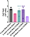
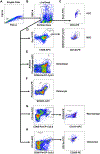
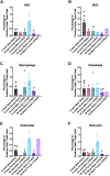
The bone microenvironment cellular composition plays an essential role in bone health and is disrupted in bone pathologies, such as osteoporosis, osteoarthritis, and cancer. Flow cytometry protocols for hematopoietic stem cell lineages are well defined and well established. Additionally, a consensus for mesenchymal stem cell flow markers has been developed. However, flow cytometry markers for bone-residing cells-osteoblasts, osteoclasts, and osteocytes-have not been proposed. Here, we describe a novel partial digestion method to separate these cells from the bone matrix and present new markers for enumerating these cells by flow cytometry. We optimized bone digestion and analyzed markers across murine, nonhuman primate, and human bone. The isolation and staining protocols can be used with either cell sorting or flow cytometry. Our method allows for the enumeration and collection of hematopoietic and mesenchymal lineage cells in the bone microenvironment combined with bone-residing stromal cells. Thus, we have established a multi-fluorochrome bone marrow cell-typing methodology. © 2022 The Authors. Current Protocols published by Wiley Periodicals LLC. Basic Protocol 1: Partial digestion for murine long bone stromal cell isolation Alternate Protocol 1: Partial digestion for primate vertebrae stromal cell isolation Alternate Protocol 2: Murine vertebrae crushing for bone stromal cell isolation Basic Protocol 2: Staining of bone stromal cells Support Protocol 1: Fluorescence minus one control, isotype control, and antibody titration Basic Protocol 3: Cell sorting of bone stromal cells Alternate Protocol 3: Flow cytometry analysis of bone stromal cells Support Protocol 2: Preparing compensation beads.
Keywords: bone marrow; cell sorting; flow cytometry; osteoblast; osteoclast; osteocyte.
© 2022 The Authors. Current Protocols published by Wiley Periodicals LLC.
Conflict of interest statement
CONFLICT OF INTEREST STATEMENT:
The authors declare there are no conflicts of interest.
Figures


References
LITERATURE CITED:
-
- Baumgarth N, and Roederer M 2000. A practical approach to multicolor flow cytometry for immunophenotyping. Journal of immunological methods 243:77–97. Available at: https://linkinghub.elsevier.com/retrieve/pii/S0022175900002295 [Accessed January 23, 2022]. - PubMed
-
- Bonewald LF 2011. The amazing osteocyte. Journal of Bone and Mineral Research 26:229–238. Available at: https://onlinelibrary.wiley.com/doi/10.1002/jbmr.320. - DOI - PMC - PubMed
-
- Boxall SA, and Jones E 2012. Markers for Characterization of Bone Marrow Multipotential Stromal Cells. Stem Cells International 2012:1–12. Available at: https://www.hindawi.com/journals/sci/2012/975871/. - PMC - PubMed
-
- Boyle WJ, Simonet WS, Lacey DL, Faust J, Hunt P, Burgess TL, Scully S, Van G, Eli A, Qian Y, et al. 1999. Osteoclast markers accumulate on cells developing from human peripheral blood mononuclear precursors. Journal of cellular biochemistry 72:67–80. - PubMed
-
- Correa D, Somoza RA, Lin P, Schiemann WP, and Caplan AI 2016. Mesenchymal stem cells regulate melanoma cancer cells extravasation to bone and liver at their perivascular niche. International journal of cancer 138:417–27. Available at: http://doi.wiley.com/10.1002/ijc.29709 [Accessed September 19, 2016]. - DOI - PMC - PubMed
INTERNET RESOURCES:
-
- https://www.stemcell.com/human-hematopoietic-stem-and-progenitor-cell-ph... A regularly updated list of phenotypic markers for progenitor cells of the hematopoietic lineage.
-
- https://www.rndsystems.com/research-area/osteoblast-and-osteoclast-markers A regularly updated list of surface and secreted markers for osteoblasts and osteoclasts.
-
- https://www.biolegend.com/en-us/blog/titrating-totalseq-antibodies A recommended antibody titration protocol.
-
- https://www.biolegend.com/Files/Images/BioLegend/literature/images/02-00... A resource describing how best to optimize multi-fluorophore flow cytometry staining.
-
- https://www.bio-rad-antibodies.com/introduction-to-flow-cytometry.html A good overview of the principles of flow cytometry with detailed explanations on preparing controls and optimizing flow data.
MeSH terms
Grants and funding
LinkOut - more resources
Full Text Sources

