. 2022 Mar 29;17(3):e0265893.
doi: 10.1371/journal.pone.0265893. eCollection 2022.

Deletion of the scavenger receptor Scarb1 in osteoblast progenitors does not affect bone mass

Affiliations

Deletion of the scavenger receptor Scarb1 in osteoblast progenitors does not affect bone mass

Michela Palmieri et al. PLoS One. .

Retraction in

Abstract

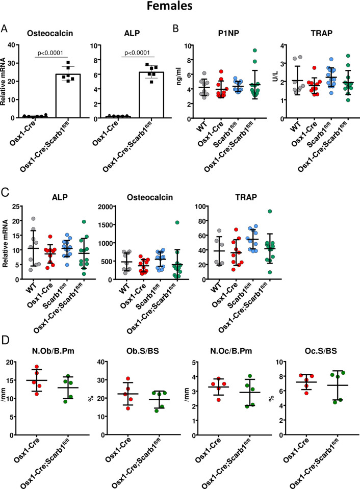
The scavenger receptor class B member 1 (SR-B1 or Scarb1) is a cell surface receptor for high density lipoproteins. It also binds oxidized low density lipoproteins and phosphocholine-containing oxidized phospholipids (PC-OxPL), which adversely affect bone homeostasis. Overexpression of a single chain form of the antigen-binding domain of E06 IgM-a natural antibody that recognizes PC-OxPL-increases trabecular and cortical bone mass in female and male mice by stimulating bone formation. We have previously reported that Scarb1 is the most abundant scavenger receptor for PC-OxPL in calvaria-derived osteoblastic cells. Additionally, bone marrow- and calvaria-derived osteoblasts from Scarb1 knockout mice (Scarb1 KO) are protected from the pro-apoptotic and anti-differentiating effects of OxPL. Previous skeletal analysis of Scarb1 KO mice has produced contradictory results, with some studies reporting elevated bone mass but another study reporting low bone mass. To clarify the role of Scarb1 in osteoblasts, we deleted Scarb1 specifically in cells of the osteoblast lineage using Osx1-Cre transgenic mice. We observed no difference in bone mineral density measured by DXA in either female or male Osx1-Cre;Scarb1fl/fl mice compared to wild type (WT), Osx1-Cre, or Scarb1fl/fl littermate controls. Additionally, microCT analysis of 6-month-old females and 7-month-old males did not detect any difference in trabecular or cortical bone mass between genotypes. These results indicate that expression of Scarb1 in cells of the osteoblast lineage does not play an important role in bone homeostasis and, therefore, it is not essential for the effects of PC-OxPL on these cells.


Conflict of interest statement

The authors have declared that no competing interests exist.

Figures

Fig 1
Fig 1. Deletion of Scarb1 increases osteoblast differentiation and protects agains the anti-proliferative effects of OxLDL.
(A) The expression of osteocalcin and alkaline phosphatase was quantified with RT-PCR in calvaria-derived osteoblasts from new born C57BL6/J (n = 5) and Scarb1 KO mice (n = 2) cultured for 21 days. Transcripts were normalized to the housekeeping gene MRPS2. Data analyzed by student t-test. (B) Proliferation was measured with Bromodeoxyuridine (BrDU) incorporation in bone marrow-derived osteoblasts from 4–5 month-old WT and Scarb1 KO mice (n = 3/group) 3 days after direct addition to the cultures of vehicle or OxLDL (50 μg/ml). Data analyzed by ANOVA, p-values were adjusted using the Holm-Sidak multiple comparison procedure. Data are shown as individual values with mean and standard deviation. All measures were performed in triplicate cultures. ALP, Alkaline phosphatase. RLU, relative light unit.
Fig 2
Fig 2. Scarb1 gene was effectively deleted in bone.
Quantitative PCR (qPCR) of loxP-flanked genomic DNA isolated from tibial cortical bone and spleen. (A) 6 month-old female mice [WT (n = 9), Osx1-Cre (n = 10), Scarb1fl/fl (n = 13), Osx1-Cre;Scarb1fl/fl (n = 12)]. (B) 7-month-old male mice [WT (n = 6), Osx1-Cre (n = 15), Scarb1fl/fl (n = 15), Osx1-Cre;Scarb1fl/fl (n = 8)].
Fig 3
Fig 3. Deletion of Scarb1 in Osx1-Cre expressing cells does not affect weight, fat or lean mass in both sexes.
(A) Weight measurements, (B) fat mass and (C) lean mass at 3 and 6 months of age in female mice [WT n = 9; Osx1-Cre n = 10; Scarb1fl/fl n = 13; Osx1-Cre;Scarb1fl/fl n = 12]. Adjusted p-values, calculated by repeated measures using two-way ANOVA are shown. (D) Weight measurements at 3 and 7 months of age in male mice [WT n = 6; Osx1-Cre n = 15; Scarb1fl/fl n = 15; Osx1-Cre;Scarb1fl/fl n = 8]. The measurements of total weight, by ANOVA repeated measures, indicated differences in the rate of weight gain between the genotypes. Further analysis, showed Scarb1fl/fl mice gained the most weight (average 11.82 ± 3.23 g) between the two time points, while WT, Osx1-Cre and Osx1-Cre; Scarb1 fl/fl gained 8.08 ± 3.46 g, 7.76 ± 3.23 g and 9.09 ± 2.22 g respectively. (E) Fat mass and (F) lean mass of the same mice as in (D) [WT n = 5–6; Osx1-Cre n = 14–15; Scarb1fl/fl n = 15; Osx1-Cre;Scarb1fl/fl n = 8]. Data are shown as mean and standard deviation.
Fig 4
Fig 4. Deletion of Scarb1 in Osx1-Cre expressing cells does not affect BMD measured by DXA.
Determinations of femoral, spinal and total BMD by DXA in: (A) 3 and 6 month old females [WT n = 9; Osx1-Cre n = 10; Scarb1fl/fl n = 13; Osx1-Cre;Scarb1fl/fl n = 12] and in (B) 3-and 7-month-old males [WT n = 5–6; Osx1-Cre n = 14–15; Scarb1fl/fl n = 15; Osx1-Cre;Scarb1fl/fl n = 8]. Data are shown as mean and standard deviation. Adjusted p-values, calculated by repeated measures using two-way ANOVA, are shown.
Fig 5
Fig 5. Deletion of Scarb1 in Osx1-Cre expressing cells does not affect trabecular bone in female mice.
Micro-CT determination of trabecular bone architecture in 6-month-old female mice in the (A) vertebra and (B) femoral metaphysis [WT n = 9; Osx1-Cre n = 10; Scarb1fl/fl n = 13; Osx1-Cre;Scarb1fl/fl n = 12]. Data are shown as mean and standard deviation. Data analyzed by ANOVA, the p-values were adjusted using the Tukey’s pairwise comparison procedure. BV/TV, bone volume /total volume. Tb, trabecular.
Fig 6
Fig 6. Deletion of Scarb1 in Osx1-Cre expressing cells does not affect trabecular bone in male mice.
Micro-CT determination of trabecular bone architecture in 7-month-old male mice in (A) vertebra [WT n = 6; Osx1-Cre n = 15; Scarb1fl/fl n = 14; Osx1-Cre;Scarb1fl/fl n = 8] and (B) femoral metaphysis [WT n = 6; Osx1-Cre n = 15; Scarb1fl/fl n = 15; Osx1-Cre;Scarb1fl/fl n = 8]. Data are shown as mean and standard deviation. Data analyzed by ANOVA, the p-values were adjusted using the Tukey’s pairwise comparison procedure. BV/TV, bone volume /total volume. Tb, trabecular.
Fig 7
Fig 7. Deletion of Scarb1 using Osx1-Cre does not affect cortical bone in female or male mice.
Micro-CT determination of cortical bone and femoral length in (A) 6-month-old female [WT n = 9; Osx1-Cre n = 10; Scarb1fl/fl n = 13; Osx1-Cre;Scarb1fl/fl n = 12], and (B) 7-month-old male mice [WT n = 6; Osx1-Cre n = 15; Scarb1fl/fl n = 15; Osx1-Cre;Scarb1fl/fl n = 8]. Data are shown as mean and standard deviation. Data analyzed by ANOVA, the p-values were adjusted using the Tukey’s pairwise comparison procedure. BV/TV, bone volume /total volume. Tb, trabecular.
Fig 8
Fig 8. Deletion of Scarb1 using Osx1-Cre increases osteoblasts differentiation in vitro, but does not affect osteoblasts or osteoclasts in vivo.
(A) The expression of osteocalcin and alkaline phosphatase was quantified with RT-PCR in calvaria-derived osteoblasts from new born Osx1-Cre (n = 5) and Osx1-Cre;Scarb1fl/fl mice (n = 5) cultured for 21 days. Transcripts were normalized to the housekeeping gene MRPS2. Data analyzed by student t-test. (B) Measurement of bone remodeling markers in the serum of 6-month-old female mice [WT n = 8; Osx1-Cre n = 10; Scarb1fl/fl n = 13; Osx1-Cre;Scarb1fl/fl n = 12], P1NP, N-terminal propeptide; TRAP, Tartrate-resistant acid phosphatase. (C) Gene expression of alkaline phosphatase, osteocalcin and TRAP in femoral cortical bone of 6-month-old female mice [WT n = 6–9; Osx1-Cre n = 10; Scarb1fl/fl n = 10–13; Osx1-Cre;Scarb1fl/fl n = 12]. Transcripts were normalized to MRPS2. Data analyzed by ANOVA, p-values >0.5. (D) Histomorphometric measurements of the trabecular vertebral bone surface (L2). [Osx1-Cre n = 5; Osx1-Cre;Scarb1fl/fl n = 5]. Data analyzed by Student t-test. N.Ob, osteoblast number. B.Pm, bone perimeter. Ob.S, osteoblast surface. BS, bone surface. N.Oc, osteoclast number. Oc.S, osteoclast surface. Data are shown as mean and standard deviation.

Similar articles

Cited by

References

    1. Shen WJ, Asthana S, Kraemer FB, Azhar S. Scavenger receptor B type 1: expression, molecular regulation, and cholesterol transport function. J Lipid Res. 2018;59(7):1114–31. doi: 10.1194/jlr.R083121 - DOI - PMC - PubMed
    1. Brundert M, Ewert A, Heeren J, Behrendt B, Ramakrishnan R, Greten H, et al.. Scavenger receptor class B type I mediates the selective uptake of high-density lipoprotein-associated cholesteryl ester by the liver in mice. Arterioscler Thromb Vasc Biol. 2005;25(1):143–8. doi: 10.1161/01.ATV.0000149381.16166.c6 - DOI - PubMed
    1. Hoekstra M, Ye D, Hildebrand RB, Zhao Y, Lammers B, Stitzinger M, et al.. Scavenger receptor class B type I-mediated uptake of serum cholesterol is essential for optimal adrenal glucocorticoid production. J Lipid Res. 2009;50(6):1039–46. doi: 10.1194/jlr.M800410-JLR200 - DOI - PMC - PubMed
    1. Zhang Y, Da Silva JR, Reilly M, Billheimer JT, Rothblat GH, Rader DJ. Hepatic expression of scavenger receptor class B type I (SR-BI) is a positive regulator of macrophage reverse cholesterol transport in vivo. J Clin Invest. 2005;115(10):2870–4. doi: 10.1172/JCI25327 - DOI - PMC - PubMed
    1. Hoekstra M, Van Eck M, Korporaal SJ. Genetic studies in mice and humans reveal new physiological roles for the high-density lipoprotein receptor scavenger receptor class B type I. Curr Opin Lipidol. 2012;23(2):127–32. doi: 10.1097/MOL.0b013e3283508c09 - DOI - PubMed

Publication types

Substances