. 2022 Aug 1;42(8):946-955.
doi: 10.1093/asj/sjac066.

Fat Grafts Augmented With Vitamin E Improve Volume Retention and Radiation-Induced Fibrosis

Affiliations

Fat Grafts Augmented With Vitamin E Improve Volume Retention and Radiation-Induced Fibrosis

Darren B Abbas et al. Aesthet Surg J. .

Abstract

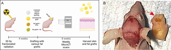
Background: Treatments for radiation-induced fibrosis range from vitamin E (VE) and pentoxifylline (PTX) systemically to deferoxamine and fat grafting locally. Regarding fat grafting, volume retention hinders its long-term functionality and is affected by 2 factors: inflammation and necrosis secondary to hypovascularity.

Objective: The authors aimed to simultaneously improve fat graft retention and radiation-induced fibrosis by integrating VE and PTX into fat grafts locally.

Methods: Forty adult CD-1 nude male mice, 6 weeks old, underwent scalp irradiation and recovered for 4 weeks to allow for development of fibrosis. Mice received 200 μL of donor human fat graft to the scalp. Mice were separated into 4 conditions: no grafting, fat graft without treatment, graft treated with PTX, and graft treated with VE. Fat graft volume retention was monitored in vivo with micro-computed tomography scans at weeks 0, 1, 2, 4, 6, and 8 after grafting. Histological and cytokine analysis of the scalp skin and fat grafts were performed.

Results: VE-treated grafts had significant improvement in dermal thickness and collagen density of overlying skin compared with all other groups. VE decreased 8-isoprostane and increased CD31+ staining compared with the other grafted groups. Cytokine analysis revealed decreased inflammatory and increased angiogenic markers in both the fat graft and overlying skin of the VE group. Fat graft volume retention was significantly improved in the VE group starting at 1 week post grafting.

Conclusions: Radiation-induced fibrosis and fat graft volume retention are both simultaneously improved with local administration of VE.


Figures

Figure 1.
Figure 1.
(A) Schematic overview of experimental design. Created with BioRender.com (Toronto, Canada). (B) Gross representation of explanted fat graft and overlying scalp skin at 8 weeks post grafting. The photograph shows the portion of the murine calvarium in direct contact with the fat graft on the left, with the explanted fat graft on the right. The graft was oriented to demonstrate the portion of the graft in direct contact with the murine calvarium (arrow), indicating proper placement of the graft within a subcutaneous pocket directly overlying the calvarium. PTX, pentoxifylline; VE, vitamin E.
Figure 2.
Figure 2.
(A) Hematoxylin and eosin (H&E)-stained sections of all conditional groups (irradiated [IR] control, untreated fat grafts [FG], FG + pentoxifylline [PTX], and FG + vitamin E [VE]) demonstrate a significant decrease in dermal thickness between the IR control cohort and all grafted groups (****P < 0.0001 for all groups). PTX and VE-treated grafts also had significantly less dermal thickening compared with the untreated FG (****P < 0.0001 for both groups). No significant difference was seen between the FG + PTX and the FG + VE cohorts regarding dermal thickness (P = 0.579). (B) Masson’s trichrome sections of all conditions. All FG groups show less collagen density compared with the IR control group (****P < 0.0001 for all groups). No significant difference was seen between the PTX and untreated FG group (P = 0.051) or between the PTX and VE groups (P = 0.149). Significantly less collagen density was seen between the untreated FG and the VE grafts (***P < 0.001). (C) Immunofluorescence imaging of 8-isoprostane staining displayed a significant decrease between IR control and untreated FG (*P < 0.05), PTX (**P < 0.01), and VE (****P < 0.0001) cohorts. The VE cohort also had significantly less 8-isoprostane staining than the untreated FG (*P < 0.05). CD31+ staining on immunofluorescence imaging demonstrated a significant increase between IR control and all grafted conditions (*P < 0.05 for untreated FG and PTX, ****P < 0.0001 for VE). VE grafts also had significantly higher CD31+ staining than untreated FG (*P < 0.05) and PTX grafts (*P < 0.05).
Figure 3.
Figure 3.
A heatmap of mouse 48-plex Luminex immunoassay (R&D Systems, Minneapolis, MN) showing cytokine levels in both fat grafts and the overlying scalp skin. The yellow and purple colors represent higher or lower levels, respectively. The vitamin E (VE) treatment group demonstrated significantly lower levels of inflammatory cytokines (interleukin-1, interleukin-2, CCL2, CCL3, CCL4, CCL7, CCL11, soluble receptor activator of nuclear factor-kappaB ligand [SRANKL], interferon-gamma [IFN-gamma]) compared with the pentoxifylline (PTX) or untreated grafts. Conversely, the vitamin E group also displayed a significantly higher level of vascular endothelial growth factor, indicating an increased amount of ongoing neovascularization both within the fat graft itself as well as the overlying skin.
Figure 4.
Figure 4.
(A) Representative images of the micro-computed tomography scans performed at the axial, coronal, and sagittal views of a vitamin E (VE)-treated graft. Utilizing this same graft, a representation of the 3-dimensional reconstructive image of the mouse calvarium with directly overlying fat graft utilized to calculate graft volume is shown. (B) A longitudinal demonstration of fat graft volume retention seen on volumetric analysis utilizing micro-computed tomography scans show a significant increase in volume retention in the vitamin E group compared with the untreated fat graft group (**P < 0.01) as early as 1 week post grafting. This effect lasted all the way until 8 weeks post grafting, where the effect is less pronounced (*P < 0.05) but still significant. The pentoxifylline (PTX) and vitamin E cohorts also show significant volumetric differences at 8 weeks post grafting (*P < 0.05).

Comment in

Similar articles

Cited by

References

    1. Deleon NMD, Adem S, Lavin CV, et al. . Angiogenic CD34+CD146+ adipose-derived stromal cells augment recovery of soft tissue after radiotherapy. J Tissue Eng Regen Med. 2021;15(12):1105-1117. doi: 10.1002/term.3253 - DOI - PMC - PubMed
    1. Borrelli MR, Deleon NMD, Adem S, et al. . Fat grafting rescues radiation-induced joint contracture. Stem Cells Dayt Ohio. 2020;38(3):382-389. doi: 10.1002/stem.3115 - DOI - PMC - PubMed
    1. Borrelli MR, Patel RA, Sokol J, et al. . Fat chance: the rejuvenation of irradiated skin. Plast Reconstr Surg Glob Open. 2019;7(2):e2092. doi: 10.1097/GOX.0000000000002092 - DOI - PMC - PubMed
    1. Lujan-Hernandez J, Chin MS, Perry DJ, et al. . Increasing fat graft retention in irradiated tissue after preconditioning with external volume expansion. Plast Reconstr Surg. 2020;145(1):103-112. doi: 10.1097/PRS.0000000000006372 - DOI - PubMed
    1. Merrifield BA, Chang A, Hostetter G, Komorowska-Timek E. Volume retention, metabolism, and cellular composition of human fat xenografts. Plast Reconstr Surg Glob Open. 2018;6(8):e1869. doi: 10.1097/GOX.0000000000001869 - DOI - PMC - PubMed