[Preprint]. 2022 Mar 24:2022.03.23.485509.
doi: 10.1101/2022.03.23.485509.

Autoantibody discovery across monogenic, acquired, and COVID19-associated autoimmunity with scalable PhIP-Seq

Affiliations

Autoantibody discovery across monogenic, acquired, and COVID19-associated autoimmunity with scalable PhIP-Seq

Sara E Vazquez et al. bioRxiv. .

Update in

  • Autoantibody discovery across monogenic, acquired, and COVID-19-associated autoimmunity with scalable PhIP-seq.
    Vazquez SE, Mann SA, Bodansky A, Kung AF, Quandt Z, Ferré EMN, Landegren N, Eriksson D, Bastard P, Zhang SY, Liu J, Mitchell A, Proekt I, Yu D, Mandel-Brehm C, Wang CY, Miao B, Sowa G, Zorn K, Chan AY, Tagi VM, Shimizu C, Tremoulet A, Lynch K, Wilson MR, Kämpe O, Dobbs K, Delmonte OM, Bacchetta R, Notarangelo LD, Burns JC, Casanova JL, Lionakis MS, Torgerson TR, Anderson MS, DeRisi JL. Vazquez SE, et al. Elife. 2022 Oct 27;11:e78550. doi: 10.7554/eLife.78550. Elife. 2022. PMID: 36300623 Free PMC article.

Abstract

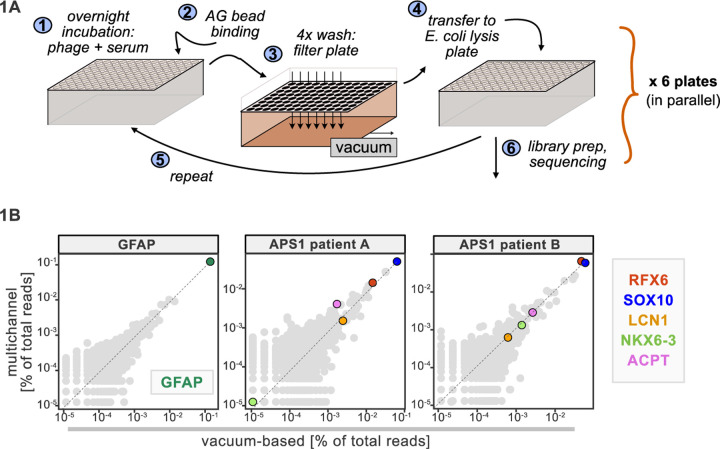
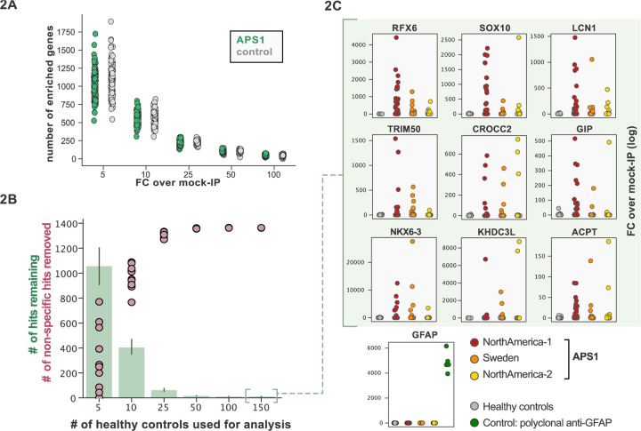
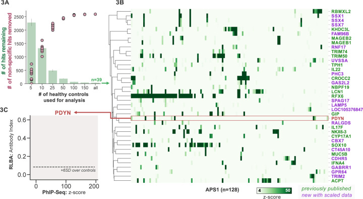
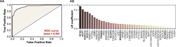
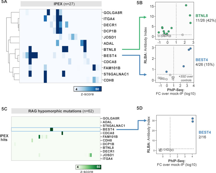
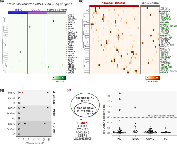
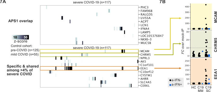
Phage Immunoprecipitation-Sequencing (PhIP-Seq) allows for unbiased, proteome-wide autoantibody discovery across a variety of disease settings, with identification of disease-specific autoantigens providing new insight into previously poorly understood forms of immune dysregulation. Despite several successful implementations of PhIP-Seq for autoantigen discovery, including our previous work (Vazquez et al. 2020), current protocols are inherently difficult to scale to accommodate large cohorts of cases and importantly, healthy controls. Here, we develop and validate a high throughput extension of PhIP-seq in various etiologies of autoimmune and inflammatory diseases, including APS1, IPEX, RAG1/2 deficiency, Kawasaki Disease (KD), Multisystem Inflammatory Syndrome in Children (MIS-C), and finally, mild and severe forms of COVID19. We demonstrate that these scaled datasets enable machine-learning approaches that result in robust prediction of disease status, as well as the ability to detect both known and novel autoantigens, such as PDYN in APS1 patients, and intestinally expressed proteins BEST4 and BTNL8 in IPEX patients. Remarkably, BEST4 antibodies were also found in 2 patients with RAG1/2 deficiency, one of whom had very early onset IBD. Scaled PhIP-Seq examination of both MIS-C and KD demonstrated rare, overlapping antigens, including CGNL1, as well as several strongly enriched putative pneumonia-associated antigens in severe COVID19, including the endosomal protein EEA1. Together, scaled PhIP-Seq provides a valuable tool for broadly assessing both rare and common autoantigen overlap between autoimmune diseases of varying origins and etiologies.


Conflict of interest statement

COMPETING INTERESTS

Figures

Figure 1.
Figure 1.. Advantages of and considerations motivating scaled PhIP-Seq.
A) Schematic of vacuum-based scaled PhIP-Seq protocol, allowing for parallelized batches of 600–800 samples. B) Comparison of moderate-throughput multichannel protocol data to high-throughput vacuum-based protocol data, with axes showing normalized read percentages. Controls include a commercial polyclonal anti-GFAP antibody (left), APS1 patient A with known and validated autoantibodies RFX6, SOX10, ACPT and LCN1 (center), and APS1 patient B with the same known and validated autoantibodies as well as NKX6–3.
Figure 2.
Figure 2.. Application of scaled PhIP-Seq to expanded APS1 and healthy control cohorts.
A) Number of hits per sample reaching 5, 10, 25, 50, and 100-fold enrichment relative to mock-IP samples. Each dot represents a single APS1 patient (green) or non-APS1 control (grey). B) When looking for disease-specific hits, increasing the number of healthy controls results in fewer apparent hits and is therefore critical. Shared hits are defined as gene-level signal (>10-fold change over mock-IP) which is shared among 10% of APS1 samples (n=128), present in fewer than 2% of healthy controls, and with at least 1 APS1 sample with a high signal (FC of 50<). Random downsampling was performed 10 times for each healthy control bin. C) 9 gene-level hits are present in 10%< of a combined 3-group APS1 cohort. North-America-1, n = 62; Sweden, n = 40; North-America-2, n = 26. Anti-GFAP control antibody (n=5) indicates that results are consistent across plates and exhibit no well-to-well contamination.
Figure 3.
Figure 3.. Replication and expansion of APS1 autoantigens across multiple cohorts using scaled PhIP-Seq.
A) Increasing the number of healthy controls results in fewer apparent hits and is therefore critical. Shared hits are defined as gene-level signal (>10-fold change over mock-IP) which is shared among 4%< of APS1 samples (n=128), present in fewer than 2% of healthy controls, and with at least 1 APS1 sample with a high signal (FC of 50<). Random downsampling was performed 10 times for each healthy control bin. B) 39 candidate hits present in 4%< of the APS1 cohort. C) Rare, novel anti-PDYN autoantibodies validate at whole-protein level, with PhIP-Seq and whole-protein RLBA data showing good concordance.
Figure 4.
Figure 4.. Logistic regression of PhIP-Seq data enables APS1 disease prediction.
A) ROC curve for prediction of APS1 versus control disease status. B) The highest logistic regression (LR) coefficients include known antigens RFX6, KHDC3L, and others.
Figure 5.
Figure 5.. PhIP-Seq screening in IPEX and RAG1/2 deficiency reveals novel, intestinally expressed autoantigens BEST4 and BTNL8.
A) PhIP-Seq heatmap of most frequent shared antigens among IPEX, with color indicating z-score relative to a cohort of non-IPEX controls. B) Radioligand binding assay for BTNL8 reveals additional anti-BTNL8 positive IPEX patients (top). Radioligand binding assay for BEST4 autoantibodies correlates well with PhIP-Seq data (bottom). C) PhIP-Seq screen of patients with hypomorphic mutations in RAG1/2 reveals 2 patients with anti-BEST4 signal. D) Orthogonal radioligand binding assay validation of anti-BEST4 antibodies in both PhIP-Seq anti-BEST4 positive patients.
Figure 6.
Figure 6.. PhIP-Seq screening of MIS-C and KD cohorts.
A) Heatmap of signal for putative hits from Gruber et al. 2020, among MIS-C, adult COVID19 controls, and pediatric febrile controls (each n=20). B) Only rare, shared PhIP-Seq signals were found among n=20 MISC patients. C) Heatmap of putative antigens in a cohort of n = 70 KD patients. Hits that are specific to KD, and are not found among n=20 febrile controls, are highlighted in green. D) A small number of rare putative antigens are shared between KD and MISC (left), with radioligand binding assay confirmation of antibody reactivity to whole protein form of CGNL1 in 3 KD patients and 1 MISC patient (right).
Figure 7.
Figure 7.. PhIP-Seq screening in severe forms of COVID-19, MIS-C and KD reveals putative novel autoantigens, including EEA1.
A) Screening of patients with severe COVID19 pneumonia shows little overlap with APS1, but enables discovery of possible novel disease associated autoantigens including EEA1. B) Putative novel antigens EEA1, CHRM5, and MCAM are primarily found in anti-IFN-negative patients, suggesting the possibility of other frequent, independent disease-associated antibodies in severe COVID19.

References

    1. Asano T., Sasaki N., Yashiro K., Hatori T., Kuwabara K., Hamada H., Imai T., and Fujino O. (2005). Acute pancreatitis with Kawasaki disease: analysis of cases with elevated serum amylase levels. Eur J Pediatr 164, 180–181. - PubMed
    1. Bacchetta R., Passerini L., Gambineri E., Dai M., Allan S.E., Perroni L., Dagna-Bricarelli F., Sartirana C., Matthes-Martin S., Lawitschka A., et al. (2006). Defective regulatory and effector T cell functions in patients with FOXP3 mutations. J Clin Invest 116, 1713–1722. - PMC - PubMed
    1. Barros R.D.M., Roberts N.A., Dart R.J., Vantourout P., Jandke A., Nussbaumer O., Deban L., Cipolat S., Hart R., Iannitto M.L., et al. (2016). Epithelia Use Butyrophilin-like Molecules to Shape Organ-Specific γδ T Cell Compartments. Cell 167, 203–218.e17. - PMC - PubMed
    1. Bastard P., Rosen L.B., Zhang Q., Michailidis E., Hoffmann H.-H., Zhang Y., Dorgham K., Philippot Q., Rosain J., Béziat V., et al. (2020). Autoantibodies against type I IFNs in patients with life-threatening COVID-19. Science 370, eabd4585. - PMC - PubMed
    1. Bastard P., Michailidis E., Hoffmann H.-H., Chbihi M., Voyer T.L., Rosain J., Philippot Q., Seeleuthner Y., Gervais A., Materna M., et al. (2021a). Auto-antibodies to type I IFNs can underlie adverse reactions to yellow fever live attenuated vaccine. J Exp Med 218. - PMC - PubMed

Publication types