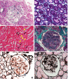
Renal Abnormalities Caused by Canine Distemper Virus Infection in Terminal Patients
- PMID: 35350433
- PMCID: PMC8957885
- DOI: 10.3389/fvets.2022.822525
Renal Abnormalities Caused by Canine Distemper Virus Infection in Terminal Patients
Abstract
The aim of this study was to analyze the glomerular and tubular alterations in dogs with terminal distemper through light microscopy, immunofluorescence, and electron microscopy. Thirteen animals with a molecular diagnosis of distemper and neurological signs were selected. As a control group, 10 clinically healthy animals with no manifestations or signs of disease and with negative tests for Ehrlichia sp., Anaplasma sp., and Babesia sp. were included in this study. Renal tissue was evaluated by light microscopy, topochemistry for DNA/chromatin, and video image analysis to detect the nuclear phenotypes of the renal tubular epithelial cells (RTECs), immunofluorescence, and transmission electron microscopy. Results showed that dogs with distemper exhibited anemia, hypergammaglobulinemia, and proteinuria. Creatinine in the distemper group was lower compared to the control group (p = 0.0026), but there was no significant difference in relation to urea (p = 0.9876). Although this alteration may be due to the smaller muscle mass observed in animals with distemper, it probably is not of clinical importance. Glomerular and tubular lesions were confirmed by light microscopy in 84.6% of these animals. Additional findings in the animals with distemper included deposition of different classes of immunoglobulins, particularly IgM in 92.3% of the cases, fibrinogen deposition in 69.2% of the cases as assessed by immunofluorescence, alterations in the nuclear phenotypes of the RTEC characterized by condensation of chromatin, loss of DNA and reduction in the nuclear shape, and the presence of subendothelial and mesangial electron-dense deposits. These findings confirm the existence of renal alterations related to terminal distemper.
Keywords: Paramyxoviridae; glomerulonephritis; glomerulosclerosis; microscopy; proteinuria; tubular necrosis.
Copyright © 2022 Silva, Silva, Borin-Crivellenti, Alvarenga, Aldrovani, Braz, Aoki, Santana, Pennacchi and Crivellenti.
Conflict of interest statement
The authors declare that the research was conducted in the absence of any commercial or financial relationships that could be construed as a potential conflict of interest.
Figures


Similar articles
-
Anosmia associated with canine distemper.Am J Vet Res. 1988 Aug;49(8):1295-7. Am J Vet Res. 1988. PMID: 3178025
-
Apoptotic investigation of brain tissue cells in dogs naturally infected by canine distemper virus.Virol J. 2021 Aug 12;18(1):165. doi: 10.1186/s12985-021-01635-8. Virol J. 2021. PMID: 34384430 Free PMC article.
-
Investigation of glomerular lesions in dogs with acute experimentally induced Ehrlichia canis infection.Am J Vet Res. 1992 Dec;53(12):2286-91. Am J Vet Res. 1992. PMID: 1476309
-
Canine distemper infections, with special reference to South Africa, with a review of the literature.J S Afr Vet Assoc. 2001 Sep;72(3):127-36. doi: 10.4102/jsava.v72i3.635. J S Afr Vet Assoc. 2001. PMID: 11811699 Review.
-
Establishment of central nervous system infection by canine distemper virus: breach of the blood-brain barrier and facilitation by antiviral antibody.Vet Immunol Immunopathol. 1987 Dec;17(1-4):471-82. doi: 10.1016/0165-2427(87)90163-2. Vet Immunol Immunopathol. 1987. PMID: 2963430 Review.
Cited by
-
Epidemiological, Clinical, and Molecular Insights into Canine Distemper Virus in the Mekong Delta Region of Vietnam.Viruses. 2025 May 29;17(6):781. doi: 10.3390/v17060781. Viruses. 2025. PMID: 40573375 Free PMC article.
-
Feline Morbillivirus: Clinical Relevance of a Widespread Endemic Viral Infection of Cats.Viruses. 2023 Oct 13;15(10):2087. doi: 10.3390/v15102087. Viruses. 2023. PMID: 37896864 Free PMC article. Review.
References
LinkOut - more resources
Full Text Sources
Research Materials

