. 2022 Mar 8:13:816498.
doi: 10.3389/fphar.2022.816498. eCollection 2022.

Moringa oleifera Seed at the Interface of Food and Medicine: Effect of Extracts on Some Reproductive Parameters, Hepatic and Renal Histology

Affiliations

Moringa oleifera Seed at the Interface of Food and Medicine: Effect of Extracts on Some Reproductive Parameters, Hepatic and Renal Histology

Alfred F Attah et al. Front Pharmacol. .

Abstract

The lipid-rich Seed of Moringa oleifera has been promoted as an effective water clarifier. Aside its vital nutritional application as an emerging food additive, the seed has continued to gain a wider acceptance in various global ethnomedicines for managing several communicable and lifestyle diseases, howbeit, its potential toxic effect, particularly on fertility and pregnancy outcomes has remained uninvestigated; the effect of Moringa oleifera seed (MOSE) aqueous-methanol extracts on fertility and pregnancy outcome, was investigated in vivo using female Wistar rats that were divided into 50, 100, 300 and 500 mg per kilogram body weight. Group six was given Moringa oleifera seed treated water ad-libitum (ad-libitum group). Organs harvested for histological assessment included ovary, uterus, liver and kidney. In addition to HPLC fingerprint and a preliminary peptide detection, we determined the physico-chemical characteristics and mineral content of MOSE using standard methods. Data were analyzed with significance at p ≤ 0.05. There was no significant difference in the estrus cycle, mating index, gestation survival index, gestation index, fertility index and sex ratio among all groups. Gestation length was reduced in some groups. While the male pup birth weight was comparable among the different groups, female pups birth weights were significantly reduced in 50 and 100 mg groups. Anogenital distance indices of female pups in ad libitum group were significantly increased. Pathologies were observed in liver and kidneys of dams while kidneys of pups presented a dose dependent reduction in the number of glomeruli. There were no observed pathological changes in the ovary and uterus. This study showed for the first time in rodents, that the lipid-rich MOSE is unsafe to the kidney of rodents while the lipid-free MOSE appears to be safe at doses up to 300 mg/kg body weight. Findings from this study suggested that the female pups were masculinized. In conclusion, the lipid-rich seed extracts of MOSE appear to be unsafe during pregnancy, induce hepatic and renal toxicity while the lipid-free MOSE excludes inherent toxicity as the hydrophobic part has been linked to toxicity as observed in this study due to the developmental programming effect on female offspring in rodents.

Keywords: Moringa oleifera; Wistar rats; developmental programming; hepatic toxicity; renal histology; reproductive parameters; seed.


Conflict of interest statement

The authors declare that the research was conducted in the absence of any commercial or financial relationships that could be construed as a potential conflict of interest.

Figures

FIGURE 1
FIGURE 1
Weight variations of Dams over the period of administration of MOSE.
FIGURE 2
FIGURE 2
Effects of Moringa oleifera Aqueous Seed Extracts on Ovarian tissue of Dams; H&E, X100, scale bar = 50 μm. Photomicrograph of an ovarian sections showing normal, developing follicles (white arrow) within the cortex. Moderate infiltration of inflammatory cells is seen in stroma (slender arrow) of 0 mg only. The stroma (slender arrows) appears normal in all other groups.
FIGURE 3
FIGURE 3
Effects of Moringa oleifera Aqueous Seed Extracts on Uterine Tissue of Dams; H&E, X100, scale bar = 50 μm. (A)—0 mg: Photomicrograph of uterus showing normal epithelial layer (white arrow) and normal endometrial glands (slender arrow). The lumen is normal. (B)—50 mg: Endometrial layer with normal epithelial layer (white arrow) and normal endometrial glands (slender arrow). The endometrium is infiltrated by inflammatory cells including lymphocytes and polymorphonuclear cells (red arrow). (C)—100 mg: Endometrium with normal epithelial layer (white arrow) and normal endometrial glands (slender arrow). The endometrium is infiltrated by inflammatory cells including lymphocytes and polymorphonuclear cells (red arrow). (D)—300 mg: Endometrial layer with normal epithelial layer (white arrow) and proliferated endometrial cells and glands (slender arrow). The endometrium is very mildly infiltrated by inflammatory cells (red arrow) including lymphocytes and polymorphonuclear cells. (E)—500 mg: Endometrial layer with normal epithelial layer (white arrow) moderately proliferated endometrial cells and glands (slender arrow) with mild infiltration of inflammatory cells (red arrow). Mild haemorrhage seen (green arrow). (F)Ad-libitum: Endometrial tissue with normal glandular cells (slender arrow) and moderate infiltration of inflammatory cells (red arrow). The epithelial linings are normal (white arrow).
FIGURE 4
FIGURE 4
Effects of Moringa oleifera Aqueous Seed Extracts on Liver of Dams; H&E, X400, scale bars = 50 μm. (A)—0 mg: Photomicrograph of normal liver section showing normal architecture, the central vessel appears normal (white arrow), the sinusoids (slender arrow) appear normal there is no infiltration. The morphology of the hepatocytes appear normal and no pathological leision is seen. (B)—50 mg: Normal liver architecture, the central venule appear normal (white arrow), no vascular congestion seen, the sinusoids show mild infiltration of inflammatory cells (slender arrow), the morphology of the hepatocytes appear normal. (C)—100 mg: Moderately normal liver architecture with normal central venule (white arrow), there are some focal areas of mild diffusion of red cells infiltrating the liver parenchyma and sinusoids (slender arrow). There is periportal infiltration (black arrow). The morphology of the hepatocytes appear normal. (D)—300 mg: Poor liver architecture. The central venule (white arrow) appear normal, no vascular congestion seen, the sinusoids appear mildly infiltrated (slender arrow). The morphology of the hepatocytes appears normal. (E)—500 mg: Poor architecture with loss of liver plate and diffused red cells (black arrow). There are hepatocytes with hyperchromic nuclei (blue arrow), others appear normal. (F)Ad-libitum: Normal liver architecture, the central venule (white arrow). No vascular congestion is seen and the sinusoids appear normal (slender arrow). The morphology of the hepatocytes appear normal.
FIGURE 5
FIGURE 5
Effects of Moringa oleifera Aqueous Seed Extracts on the Kidney of Dams; H&E, X400, scale bars = 50 μm. (A)—0 mg: photomicrograph of normal kidney section showing normal renal cortex with normal glomeruli (white arrow) containing normal mesangial cells and capsular spaces. The renal tubules shows normal epithelial distribution and appear normal (blue arrow), the interstitial spaces (slender arrow) are not infiltrated. (B)—50 mg: Some glomeruli with mild mesangial cells hyperplasia (white arrow). The renal tubules show mild tubular necrosis and eosinophilic cast (black arrow) within their lumen. The interstitial spaces are midly infiltrated (slender arrow). (C)—100 mg: Normal glomeruli containing normal mesangial cells and capsular spaces (white arrow). The renal tubules show moderate tubular necrosis with loss of brush border (blue arrow). There is very mild infiltration of interstitial spaces (slender arrow). (D)—300 mg: Normal glomeruli containing normal mesangial cells and capsular spaces (white arrow). The renal tubules shows moderate tubular necrosis with loss of brush border (blue arrow) while other tubules are intact (black arrow). The interstitial spaces appear normal (slender arrow). (E)—500 mg: Tubules with moderate tubular necrosis (blue arrow). The interstitial spaces show scanty infiltration (slender arrow). (F)Ad-libitum: Normal glomeruli containing normal mesangial cells and capsular spaces (white arrow). There is mild tubular necrosis (blue arrow) while other renal tubules appear normal (black arrow).
FIGURE 6
FIGURE 6
Effects of Moringa oleifera Aqueous Seed Extracts on the Kidney of Pup; H&E, X400, scale bars = 50 μm. (A)—0 mg: Photomicrograph of normal kidney section showing normal multiple renal cortex with normal glomeruli (white arrow) containing normal mesangial cells and capsular spaces. The interstitial spaces show mild infiltration (slender arrow). (B)—50 mg: Numerous normal glomeruli within the renal cortex (white arrow). The renal tubules show attenuation and some tubules appear collapsed with lack of luminar spaces (black arrow). The interstitial spaces appear normal (slender arrow). (C)—100 mg: Numerous normal glomeruli within the renal cortex (white arrow). Some tubules are collapsed with diminished lumen (blue arrow) while others appear normal. The interstitial space is moderately widened and infiltrated by red cells (slender arrow) and few inflammatory cells. (D)—300 mg: Numerous compact glomeruli within the renal cortex (white arrow). The renal tubules shows acute tubular necrosis with loss of brush border (blue arrow). There are limiting lumen in the tubules as some tubules appear collapsed, the interstitial space is hardly defined (slender arrow). (E)—500 mg: Compact glomeruli within the renal cortex (white arrow). The renal parenchyma shows moderate fibrosis and tubules shows mild tubular necrosis (blue arrow). Some tubules are collapsed. There is vascular congestion seen in the interstitium (slender). The interstitial space is mildly infiltrated. (F)Ad-libitum: Normal glomeruli containing normal mesangial cells and capsular spaces (white arrow). The renal tubules appear normal (blue arrow), the interstitial spaces appear normal and not infiltrated (slender arrow).
FIGURE 7
FIGURE 7
Effects of Moringa oleifera Lam. Aqueous Seed Extracts on the Liver of Pup; H&E, X100, scale bars = 50 μm. (A)—0 mg: Photomicrograph of normal liver section showing normal architecture, the central vessel appear normal (white arrow), the sinusoids appear normal (slender arrow) and not infiltrated. No pathological leision seen. (B)—50 mg: Poor liver architecture. There is moderate vascular congestion seen (black arrow). The sinusoids show no infiltration of inflammatory cells (slender arrow). (C)—100 mg: Moderately normal liver architecture with normal central venule (white arrow). There is mild focal area of mild diffusion of red cells and inflammatory cells infiltrating the liver parenchyma and sinusoids (slender arrow). (D)—300 mg: Central venule (white arrow) without congestion. The sinusoids arewide and mildly infiltrated (slender arrow). (E)—500 mg: Poor liver architecture. The venule and portal vein are congested (white arrow), the sinusoids show moderate dilatation with mild infiltration of inflammatory cells, (slender arrow). (F)Ad-libitum: Normal liver architecture. There is mild vascular congestion seen (blue arrow), the sinusoids appear normal with no infiltration.
FIGURE 8
FIGURE 8
Effect of administration of 300 mg/kg body weight MOSE Cake freed of oil on some selected organs of Wistar rats; H&E, X100, scale bar = 50 μm. (A) Photomicrographs of kidney sections showing moderately normal architecture, the renal cortex shows normal glomeruli with normal capsular spaces and mesengial cells (white arrow), most the renal tubules appear normal (blue arrow), however, few area s with collapsed tubules are noted (red arrow). the interstitial spaces appear normal and not infiltrated (slender arrow). (B) Moderate architecture of the central venules and portal tracts are not congested (white arrows), the sinusoids show scanty infiltration of inflammatory cells (slender arrow). Hepatocytes show normal morphology (blue arrow). (C) Normal uterus, the epithelium (spanned) of the uterine mucosa layer (endometrium) appears normal (white arrow), the myometrium and perimetrium appear normal, the lumen is clean (red arrow), the uterine glands show mild dilatation (blue arrow) (blue arrow).
FIGURE 9
FIGURE 9
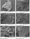
Morphology of MOSE as seed-cake powder (images ACE) and whole seed powder (images BDE). Mineral, heavy metal and RP-HPLC fingerprint of MOSE aqueous extract.
FIGURE 10
FIGURE 10
RP-HPLC chromatogram showing a general fingerprint and chemical nature of the prepurified aqueous extract of MOSE; mainly indicating early and late early eluting metabolites as expected which have been preliminarily analyzed by MALDI-TOF MS (Koehbach et al., 2013) to contain abundant peptide fragments (ranging from 1000 Da to 4.0 kDa within the mass window analyzed).

References

    1. Aboagye G., Navele M., Essuman E. (2021). Protocols for Assessing Antibacterial and Water Coagulation Potential of Moringa Oleifera Seed Powder. MethodsX 8, 101283. 10.1016/j.mex.2021.101283 - DOI - PMC - PubMed
    1. Ajayi A. F., Akhigbe R. E. (2020). Staging of the Estrous Cycle and Induction of Estrus in Experimental Rodents: an Update. Fertil. Res. Pract. 6 (1), 5–15. 10.1186/s40738-020-00074-3 - DOI - PMC - PubMed
    1. Al-Anizi A. A., Hellyer M. T., Zhang D. (2014). Toxicity Assessment and Modelling of Moringa Oleifera Seeds in Water Purification by Whole Cell Bioreporter. Water Res. 56, 77–87. 10.1016/j.watres.2014.02.045 - DOI - PubMed
    1. Alhimaidi A. R., Ammari A. A., Alghadi M. Q., Al Saiady M. Y., Amran R. A., Swelum A. A. (2021). Sex Preselection of Sٍheep Embryo by Altering the Minerals of Maternal Nutrition. Saudi J. Biol. Sci. 28 (1), 680–684. 10.1016/j.sjbs.2020.10.059 - DOI - PMC - PubMed
    1. Ashour E. A., El-Kholy M. S., Alagawany M., Abd El-Hack M. E., Mohamed L. A., Taha A. E., et al. (2020). Effect of Dietary Supplementation with Moringa Oleifera Leaves And/or Seeds Powder on Production, Egg Characteristics, Hatchability and Blood Chemistry of Laying Japanese Quails. Sustainability 12 (6), 2463. 10.3390/su12062463 - DOI

LinkOut - more resources