. 2022 Mar;12(3):210341.
doi: 10.1098/rsob.210341. Epub 2022 Mar 30.

Killing the competition: a theoretical framework for liver-stage malaria

Affiliations

Killing the competition: a theoretical framework for liver-stage malaria

Clemente F Arias et al. Open Biol. 2022 Mar.

Abstract

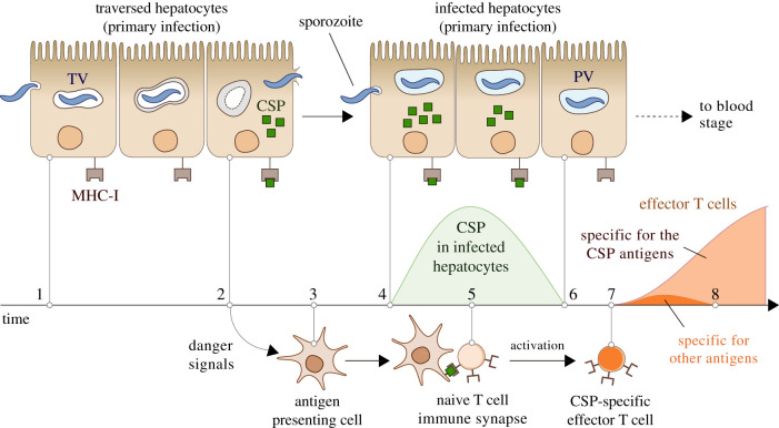
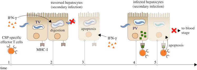
The first stage of malaria infections takes place inside the host's hepatocytes. Remarkably, Plasmodium parasites do not infect hepatocytes immediately after reaching the liver. Instead, they migrate through several hepatocytes before infecting their definitive host cells, thus increasing their chances of immune destruction. Considering that malaria can proceed normally without cell traversal, this is indeed a puzzling behaviour. In fact, the role of hepatocyte traversal remains unknown to date, implying that the current understanding of malaria is incomplete. In this work, we hypothesize that the parasites traverse hepatocytes to actively trigger an immune response in the host. This behaviour would be part of a strategy of superinfection exclusion aimed to reduce intraspecific competition during the blood stage of the infection. Based on this hypothesis, we formulate a comprehensive theory of liver-stage malaria that integrates all the available knowledge about the infection. The interest of this new paradigm is not merely theoretical. It highlights major issues in the current empirical approach to the study of Plasmodium and suggests new strategies to fight malaria.

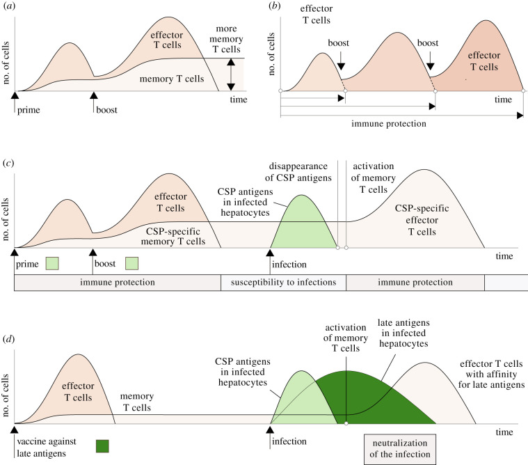
Keywords: Plasmodium; antimalarial vaccines; concomitant immunity; liver-stage malaria; superinfection exclusion.


Conflict of interest statement

We declare we have no competing interests.

Figures

Figure 1.
Figure 1.
Hypothesized mechanism of Plasmodium’s immune evasion during liver-stage malaria. Hepatocyte traversal starts after the sporozoites cross the sinusoidal barrier (1). The digestion of the transient vacuole (TV) and the cell damage caused by migrating sporozoites trigger in traversed hepatocytes the release of danger signals and the initiation of an inflammatory reaction (2) that attracts innate immune cells to the site of the infection (3). To infect hepatocytes, the parasite makes extensive use of the CSP, which leads to a significant display of CSP antigens on the membrane of host cells (4). Simultaneously, innate immune cells start the activation of T-cell clones with affinity for those antigens, a process that involves the formation of immune synapses that may last several hours (5). When the parasite takes control of the infected cell, the levels of the CSP drop to undetectable levels (6). This occurs before the activation of T cells is complete (7). In the absence of CSP antigens on their membranes, infected host cells are effectively invisible to the CSP-specific effector T cells that reach the liver (8). The immunodominance of the CSP antigens would also diminish the expansion of T-cell clones with affinity for other non-dominant sporozoite antigens that might appear in later stages of the infection [93], further contributing to the invisibility of infected hepatocytes.
Figure 2.
Figure 2.
Hypothesized mechanism of immune susceptibility of secondary Plasmodium sporozoites. The immune reaction activated by a primary infection in the host’s liver results in the production of interferon (IFN)-γ and the activation of effector T cells with affinity for the CSP (1). IFN-γ activates the digestive machinery of traversed hepatocytes, increasing the risk of sporozoite destruction in the transient vacuole (TV) (2). The cell damage inflicted by migrating sporozoites could also lead to apoptosis in traversed hepatocytes, a reaction fostered by IFN-γ that would entail the death of the parasite (3). Some sporozoites may survive hepatocyte traversal (4) and infect hepatocytes. As occurs during primary infections, secondary parasites must use the CSP in the early stages of hepatocyte invasion, which leads to the large display of CSP antigens on the membrane of newly infected host cells (4). In contrast to primary infections, CSP-specific effector T cells are already present in the liver at this moment. This increases the probability of detection and destruction of infected hepatocytes before the disappearance of the CSP from their cytoplasm, which prevents the onset of the blood stage of the infection (5).
Figure 3.
Figure 3.
Dynamics of the interactions between Plasmodium sporozoites and the T-cell response of the host. (a) The presence of the parasite in the liver triggers an adaptive immune response (1) that leads to the activation of T cells that recognize sporozoite antigens (2). This response cannot stop the progression of the infection (figure 1) but prevents the establishment of secondary sporozoites in the liver (3) (figure 2). The immune protection provided by effector T cells disappears after clonal contraction, making the host vulnerable to new infections (reinfections). In case of reinfection (4), the adaptive response to sporozoites causes the activation of memory T cells in the liver (5). However, the host’s immune memory cannot neutralize the infection and avoid the onset of blood-stage malaria. (b) We hypothesize that the number of memory T cells does not improve the immune protection of the host. The immune reaction triggered by the sporozoites in the liver (1) attracts immune cells to the site of the infection. A greater number of memory T cells in the liver increases the rate of encounter with antigen presenting cells, accelerating the start of T-cell activation (2 versus 3). By contrast, the duration of the immune synapse (several hours) cannot be shortened by increasing the number of memory T cells (4 and 5). Thus, even if more memory T cells exhibit a greater clonal expansion (6), the appearance of effector T cells is necessarily delayed with respect to the entry of the parasite in the liver. (c) For this reason, infected hepatocytes could no longer display CSP antigens after memory T-cell activation. Therefore, activated memory T cells would not stop the reinfection even though they protect the host against future secondary infections.
Figure 4.
Figure 4.
Hypothesized interactions between Plasmodium sporozoites and antimalarial vaccines. (a) In prime-boost protocols, a first dose of the vaccine triggers an immune reaction in the host and later inoculations boost this response, leading to the appearance of more memory T cells. (b) We hypothesize that the protection of prime-boost vaccines is not mediated by the formation of more memory T cells but by their effect on the duration of the T-cell response. Successive inoculations would extend the presence of effector T cells in the liver and consequently the protection against new infections (figure 2). (c) Vaccines targeting early antigens (those that disappear from infected hepatocytes within a few hours of the entry of the parasite) only confer the host with a transient immune protection. In the case of infection, those antigens are no longer present in infected hepatocytes after the activation of liver-resident memory T cells. This makes infected hepatocytes undetectable to the immune memory created by the vaccine. Activated memory T cells contribute to the strategy of superinfection exclusion of the parasite by preventing secondary infections in the host. (d) Targeting late antigens would protect the host against malaria infections. In this case, the activation of the memory T cells formed by the vaccine would coincide with the expression of target antigens on infected hepatocytes, increasing the probability of an effective neutralization of the infection.

Similar articles

Cited by

References

    1. Medica DL, Sinnis P. 2005. Quantitative dynamics of Plasmodium yoelii sporozoite transmission by infected anopheline mosquitoes. Infect. Immun. 73, 4363-4369. (10.1128/IAI.73.7.4363-4369.2005) - DOI - PMC - PubMed
    1. Gomes PS, Bhardwaj J, Rivera-Correa J, Freire-De-Lima CG, Morrot A. 2016. Immune escape strategies of malaria parasites. Front. Microbiol. 7, 1617. (10.3389/fmicb.2016.01617) - DOI - PMC - PubMed
    1. Vanderberg JP, Sidjanski S. 1997. Delayed migration of Plasmodium sporozoites from the mosquito bite site to the blood. Am. J. Trop. Med. Hyg. 57, 426-429. (10.4269/ajtmh.1997.57.426) - DOI - PubMed
    1. Protzer U, Maini MK, Knolle PA. 2012. Living in the liver: hepatic infections. Nat. Rev. Immunol. 12, 201-213. (10.1038/nri3169) - DOI - PubMed
    1. Usynin I, Klotz C, Frevert U. 2007. Malaria circumsporozoite protein inhibits the respiratory burst in Kupffer cells. Cell. Microbiol. 9, 2610-2628. (10.1111/j.1462-5822.2007.00982.x) - DOI - PubMed

Publication types