. 2022 Mar 29;14(1):26.
doi: 10.1186/s11689-022-09431-3.

Computational analysis of cortical neuronal excitotoxicity in a large animal model of neonatal brain injury

Affiliations

Computational analysis of cortical neuronal excitotoxicity in a large animal model of neonatal brain injury

Panagiotis Kratimenos et al. J Neurodev Disord. .

Abstract

Background: Neonatal hypoxic brain injury is a major cause of intellectual and developmental disability. Hypoxia causes neuronal dysfunction and death in the developing cerebral cortex due to excitotoxic Ca2+-influx. In the translational piglet model of hypoxic encephalopathy, we have previously shown that hypoxia overactivates Ca2+/Calmodulin (CaM) signaling via Sarcoma (Src) kinase in cortical neurons, resulting in overexpression of proapoptotic genes. However, identifying the exact relationship between alterations in neuronal Ca2+-influx, molecular determinants of cell death, and the degree of hypoxia in a dynamic system represents a significant challenge.

Methods: We used experimental and computational methods to identify molecular events critical to the onset of excitotoxicity-induced apoptosis in the cerebral cortex of newborn piglets. We used 2-3-day-old piglets (normoxic [Nx], hypoxic [Hx], and hypoxic + Src-inhibitor-treatment [Hx+PP2] groups) for biochemical analysis of ATP production, Ca2+-influx, and Ca2+/CaM-dependent protein kinase kinase 2 (CaMKK2) expression. We then used SimBiology to build a computational model of the Ca2+/CaM-Src-kinase signaling cascade, simulating Nx, Hx, and Hx+PP2 conditions. To evaluate our model, we used Sobol variance decomposition, multiparametric global sensitivity analysis, and parameter scanning.

Results: Our model captures important molecular trends caused by hypoxia in the piglet brain. Incorporating the action of Src kinase inhibitor PP2 further validated our model and enabled predictive analysis of the effect of hypoxia on CaMKK2. We determined the impact of a feedback loop related to Src phosphorylation of NMDA receptors and activation kinetics of CaMKII. We also identified distinct modes of signaling wherein Ca2+ level alterations following Src kinase inhibition may not be a linear predictor of changes in Bax expression. Importantly, our model indicates that while pharmacological pre-treatment significantly reduces the onset of abnormal Ca2+-influx, there exists a window of intervention after hypoxia during which targeted modulation of Src-NMDAR interaction kinetics in combination with PP2 administration can reduce Ca2+-influx and Bax expression to similar levels as pre-treatment.

Conclusions: Our model identifies new dynamics of critical components in the Ca2+/CaM-Src signaling pathway leading to neuronal injury and provides a feasible framework for drug efficacy studies in translational models of neonatal brain injury for the prevention of intellectual and developmental disabilities.

Keywords: Calcium/calmodulin; Computational modeling; Excitotoxicity; Neonatal brain injury; Nuclear calcium; SimBiology; Src kinase.


Conflict of interest statement

The authors declare that they have no competing interests.

Figures
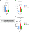
Fig. 1
Fig. 1
Experimental analysis of biochemical correlates and CaMKK2 expression in the piglet model of neonatal hypoxic brain injury. a ATP measurement in cerebral cortex tissue from piglets in the Nx (blue), Hx (red), and Hx+PP2 (green) groups; statistics: one-way ANOVA overall: F (2, 10) = 89.02 (P < 0.0001), Tukey multiple comparison test: Nx vs. Hx: P < 0.0001, Hx vs. Hx+PP2: P = 0.9253, Nx vs. Hx+PP2: P < 0.0001. b Nuclear Ca2+ influx in cerebral cortical tissue for all groups; statistics: one-way ANOVA overall: F (2, 14) = 32.94 (P < 0.0001), Tukey multiple comparison test: Nx vs. Hx: P < 0.0001, Hx vs. Hx+PP2: P = 0.0116, Nx vs. Hx+PP2: P = 0.0007. c Western blots of CaMKK2 for Nx, Hx, and Hx+PP2 groups, control: β-actin. d Quantification of CaMKK2 protein expression from western blots; statistics: one-way ANOVA overall: F (2, 15) = 19.68 (P < 0.0001), Tukey multiple comparison test: Nx vs. Hx: P = 0.0004, Hx vs. Hx+PP2: P = 0.0002, Nx vs. Hx+PP2: P = 0.9336
Fig. 2
Fig. 2
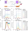
Model input. a Glutamate pulses during the three stages of the simulation—normoxia (Nx), hypoxia (Hx), and Hx+PP2. b Amplified view of Nx glutamate stimulation and c amplified view of Hx and Hx+PP2
Fig. 3
Fig. 3
Ca2+/CaM-Src kinase model validation. a Cartoon schematic of Src kinase activation of the Ca2+/CaM signaling cascade and the pro-apoptotic protein—Bax—following hypoxic neonatal brain injury, as well as pharmacological inhibition of this pathway using PP2. b Simulation of our model yields activated Src dynamics c activated CaMKK2 (blue) and CaMKIV (cyan), d cytoplasmic (maroon) and nuclear Ca2+ (grey), ATP (purple), and e Bax (black) over simulation time course
Fig. 4
Fig. 4
Global sensitivity analysis (GSA) of the Ca2+/CaM-Src model. a Input parameters include “stim” (normal glutamate pulse concentration), “hyperstim” (excitotoxic glutamate pulse concentration), NMDAR, CREB, and nuclear CaMKIV, with apoptosis (Bax) and Ca2+ influx as model readouts. b First-order Sobol indices for Bax expression. c Total-order Sobol indices for Bax expression. d First-order Sobol indices for nuclear Ca2+ influx. e Total-order Sobol indices for nuclear Ca2+ influx
Fig. 5
Fig. 5
Multi-parametric global sensitivity analysis (MPGSA) of the Ca2+/CaM-Src model. a All simulation runs (dotted grey lines) over time course; mean model simulation response (red), and the 90% region (blue) for all simulation runs for nuclear Ca2+ influx (top) and Bax expression (bottom). b Cumulative distribution function plots for MPGSA with classifier max(nuclear Ca2+) > 10 μM (left) and max(Bax expression) > 1 μM (right), showing accepted (blue) and rejected (red) sample simulation values. Asterisks represents P < 0.0001 for Kolmogorov-Smirnov test to identify difference between accepted and rejected CDFs. [Statistics: (max(Bax) > 1 μM: CaMKIV: K-S: 0.0247; P = 0.9991; CREB: K-S: 0.4137; P < 0.0001; NMDAR: P = 0.9992, K-S: 0.0246; hyperstim: P < 0.0001; K-S: 0.7736; stim: P > 0.05; K-S: 0.0193; max(nuclear Ca2+ > 10 μM): CaMKIV: P > 0.05, K-S: 0.0139; CREB: P > 0.05, K-S = 0.0136; NMDAR: P > 0.05, K-S = 0.0153; hyperstim: P < 0.0001, K-S = 1; stim: P > 0.05, K-S = 0.0104]
Fig. 6
Fig. 6
Parameter scanning to identify the relative dependence of signaling components. a Cartoon schematic showing Src-mediated NMDAR activation kinetics in comparison to CaMKII activation kinetics. b Dependence of NMDAR hyperstimulating activation kinetics on simulation outcomes for Ca2+ influx and c Bax expression. d Parameter scanning of CaMKII activation kinetics on simulation outcomes for Ca2+ influx and e Bax expression
Fig. 7
Fig. 7
Effect of PP2 pre-treatment versus combination of PP2 administration following Hx and modulation of Src kinase activation of NMDARs (Ka). a Cartoon schematic showing the effect of Src on NMDAR activation in relation to pharmacological treatment timeline. b Comparison of PP2-pre-treatment (dotted line) versus Ka = 0.01 (light tone blue), Ka = 0.012 (mid-tone blue), Ka = 0.014 (blue) on cytoplasmic Ca2+ influx and c Bax expression

Similar articles

Cited by

References

    1. Burd I, Welling J, Kannan G, Johnston M, v. Excitotoxicity as a common mechanism for fetal neuronal injury with hypoxia and intrauterine inflammation. Adv Pharmacol. 2016;76:85–101. - PubMed
    1. Northington FJ, Chavez-Valdez R, Martin LJ. Neuronal cell death in neonatal hypoxia-ischemia. Ann Neurol. 2011;69:743–758. - PMC - PubMed
    1. Lawn JE, Cousens S, Zupan J. 4 million neonatal deaths: When? Where? Why? Lancet. 2005;365:891–900. - PubMed
    1. Rossi DJ, Oshima T, Attwell D. Glutamate release in severe brain ischaemia is mainly by reversed uptake. Nature. 2000;403(6767):316–321. - PubMed
    1. Abdel-Hamid KM, Tymianski M. Mechanisms and effects of intracellular calcium buffering on neuronal survival in organotypic hippocampal cultures exposed to anoxia/aglycemia or to excitotoxins. J Neurosci. 1997;17:3538–3553. - PMC - PubMed

Publication types

Substances