Review
. 2022 May;298(5):101867.
doi: 10.1016/j.jbc.2022.101867. Epub 2022 Mar 26.

A primer to traction force microscopy

Affiliations
Review

A primer to traction force microscopy

Andrea Zancla et al. J Biol Chem. 2022 May.

Abstract

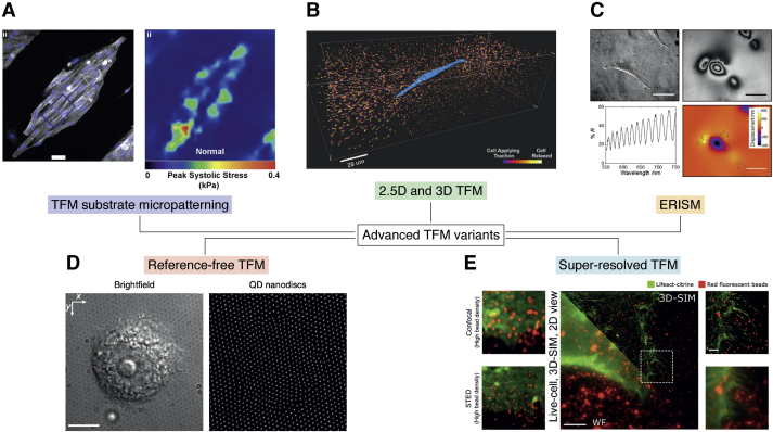
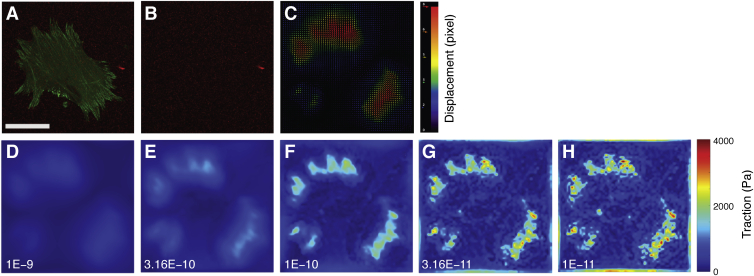
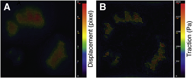
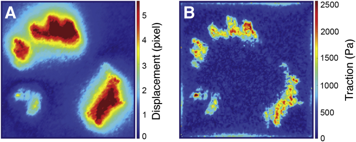
Traction force microscopy (TFM) has emerged as a versatile technique for the measurement of single-cell-generated forces. TFM has gained wide use among mechanobiology laboratories, and several variants of the original methodology have been proposed. However, issues related to the experimental setup and, most importantly, data analysis of cell traction datasets may restrain the adoption of TFM by a wider community. In this review, we summarize the state of the art in TFM-related research, with a focus on the analytical methods underlying data analysis. We aim to provide the reader with a friendly compendium underlying the potential of TFM and emphasizing the methodological framework required for a thorough understanding of experimental data. We also compile a list of data analytics tools freely available to the scientific community for the furtherance of knowledge on this powerful technique.

Keywords: biophysics; cell adhesion; cytoskeleton; focal adhesion; mechanosignaling; mechanotransduction; traction force microscopy.


Conflict of interest statement

Conflict of interest The authors declare that they have no conflicts of interest with the contents of this article.

Figures

Figure 1
Figure 1
Simplified schematic representation of the focal adhesions. Following integrin contact with the extracellular matrix (ECM), docking proteins are recruited that transfer the external stimuli toward the cytoskeleton, where traction forces are generated by the interaction between filamentous actin (F-actin) fibers and the motor protein myosin.
Figure 2
Figure 2
Advanced variants of TFM technique. Image thumbnails have been adapted with permission from the corresponding references. A, TFM substrate micropatterning (40). B, 2.5D and 3D TFM (44). C, elastic resonator interference stress microscopy (ERISM) (52). D, reference-free TFM (54). E, super-resolved TFM (55, 58). QD, quantum dot; SIM, structured illumination microscopy; STED, STimulated Emission Depletion; TFM, traction force microscopy.
Figure 3
Figure 3
Effect of the chosen regularization parameter on the estimation of the traction force magnitude. A, sample cell, expressing a fluorescent variant of the FA-associated protein paxillin (in green) and adherent to a 15-kPa (shear modulus) polyacrylamide gel laden with fluorescent nanoparticles (in red). Scale bar: 50 μm. B, reference image of the same ROI after cell detachment by trypsinization. Datasets were processed using PIV/FTTC plugins for ImageJ (see BOX 2 for details on software tools), and the resulting traction maps were post-processed in MATLAB (MathWorks, R2019b) for visualization. C, displacement vector field calculated by PIV algorithm. D-H, traction maps obtained from the displacement field in (C) using FTTC plugin with different values for the regularization parameter (in the range 1E-9 – 1E-11). The same colormap value ranges were used for all traction maps, emphasizing the impact of the chosen regularization parameter on the resulting solution. FA, focal adhesion; FTTC, Fourier transform traction cytometry; PIV, particle image velocimetry; ROI, range of interest.
Figure 4
Figure 4
Output of TFM analysis performed by PIV/FTTC plugins for ImageJ. Source dataset is as in Figure 3. A, vector plot of the displacement field (in pixels). B, vector plot of the cell traction field (in Pa). FTTC, Fourier transform traction cytometry; PIV, particle image velocimetry; TFM, traction force microscopy.
Figure 5
Figure 5
Output of Danuser Lab TFM package in MATLAB. Source dataset is as in Figure 3. A, displacement map (in pixels). B, traction intensity map (in Pa). TFM, traction force microscopy

References

    1. Iskratsch T., Wolfenson H., Sheetz M.P. Appreciating force and shape — the rise of mechanotransduction in cell biology. Nat. Rev. Mol. Cell Biol. 2014;15:825–833. - PMC - PubMed
    1. Wehrle-Haller B. Structure and function of focal adhesions. Curr. Opin. Cell Biol. 2012;24:116–124. - PubMed
    1. Schiller H.B., Fässler R. Mechanosensitivity and compositional dynamics of cell–matrix adhesions. EMBO Rep. 2013;14:509–519. - PMC - PubMed
    1. Hytönen V.P., Wehrle-Haller B. Mechanosensing in cell–matrix adhesions – converting tension into chemical signals. Exp. Cell Res. 2016;343:35–41. - PubMed
    1. del Rio A., Perez-Jimenez R., Liu R., Roca-Cusachs P., Fernandez J.M., Sheetz M.P. Stretching single talin rod molecules activates vinculin binding. Science. 2009;323:638–641. - PMC - PubMed

Publication types

LinkOut - more resources