. 2022 May 4;30(5):2092-2107.
doi: 10.1016/j.ymthe.2022.03.016. Epub 2022 Mar 26.

Host liver-derived extracellular vesicles deliver miR-142a-3p induces neutrophil extracellular traps via targeting WASL to block the development of Schistosoma japonicum

Affiliations

Host liver-derived extracellular vesicles deliver miR-142a-3p induces neutrophil extracellular traps via targeting WASL to block the development of Schistosoma japonicum

Lifu Wang et al. Mol Ther. .

Abstract

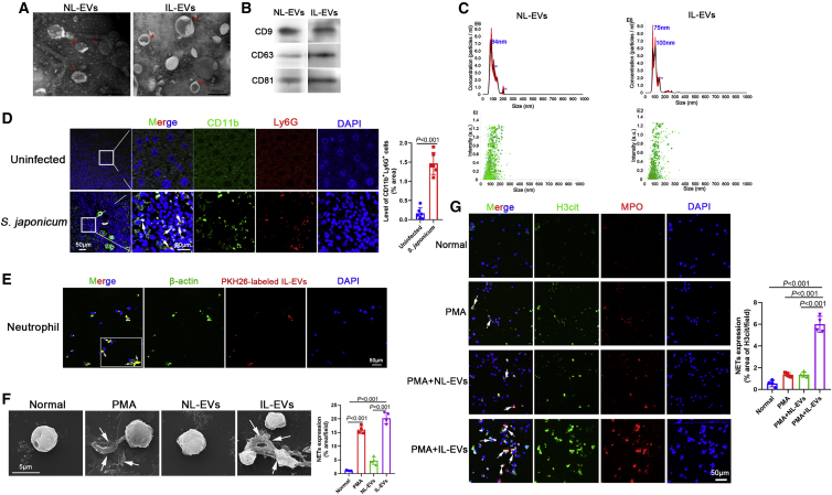
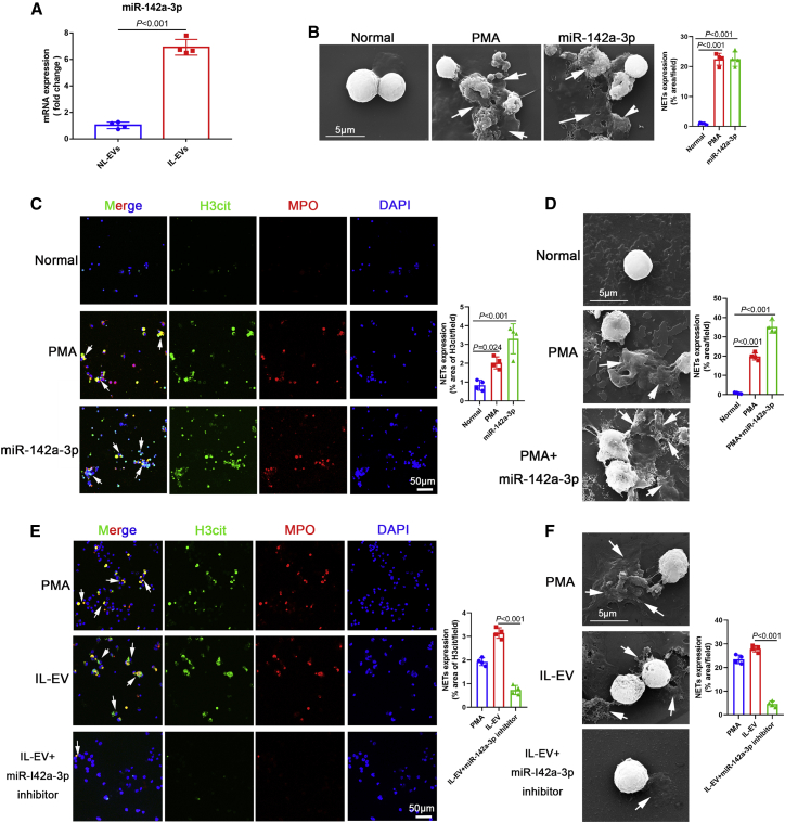
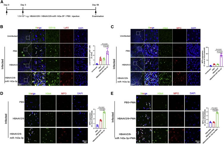
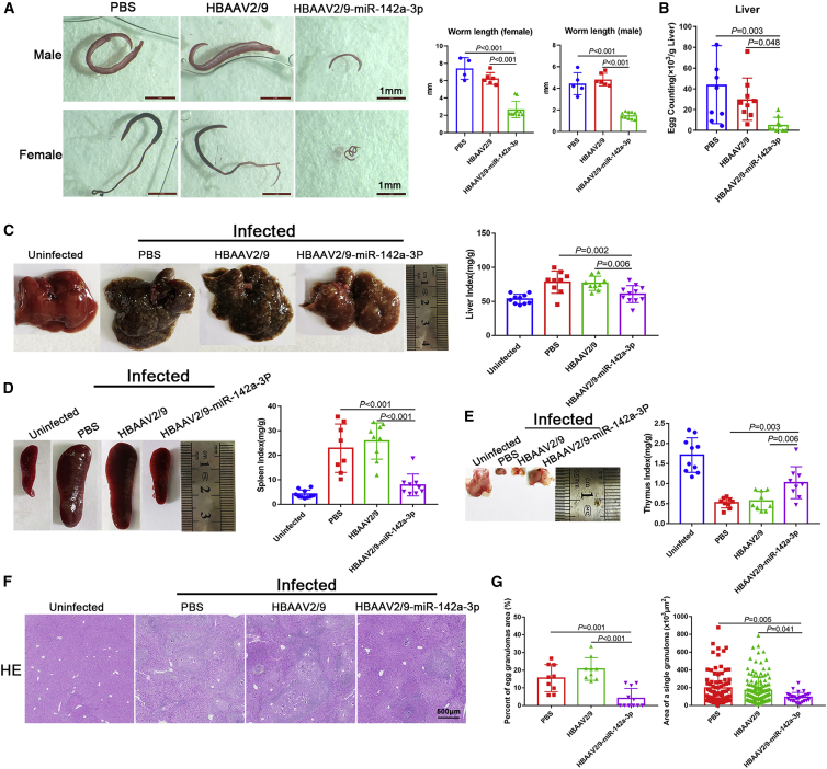
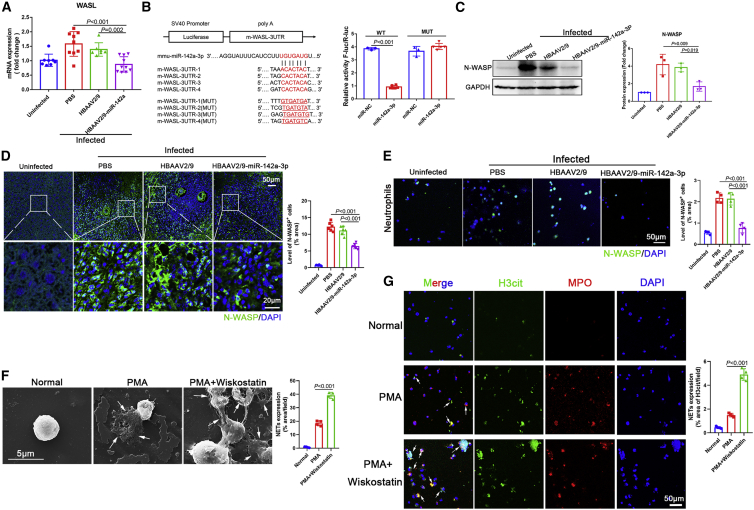
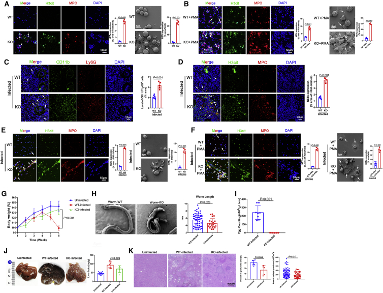
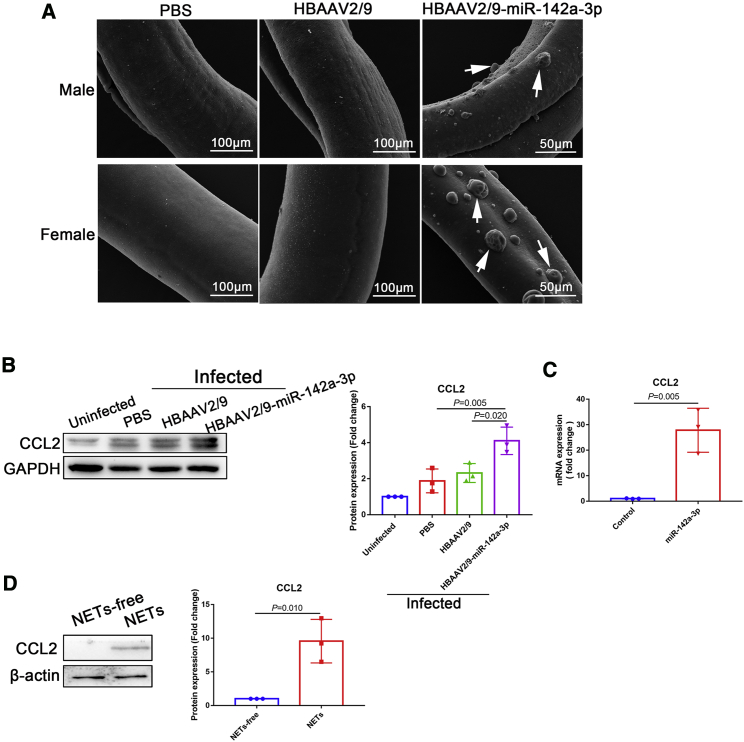
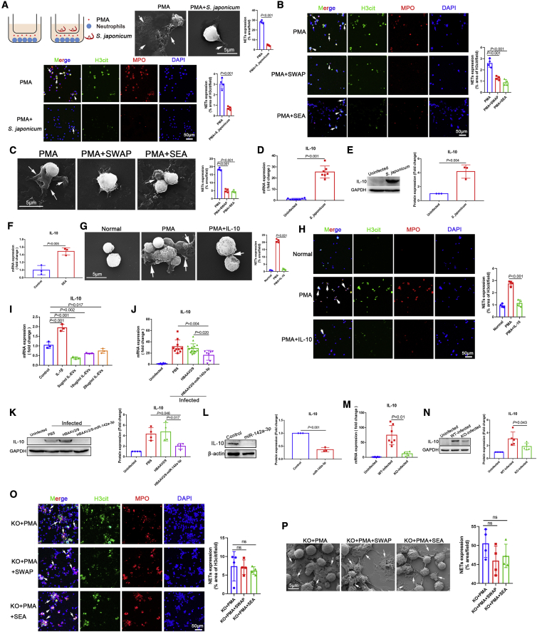
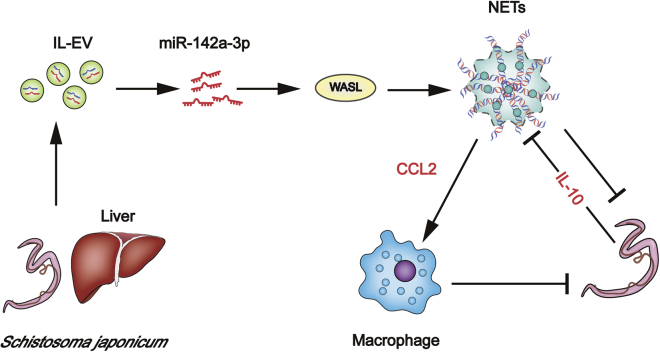
Schistosomiasis is an important neglected tropical disease. Interactions between the host immune system and schistosomes are complex. Neutrophils contribute to clearance of large pathogens primarily by releasing neutrophil extracellular traps (NETs). However, the functional role of NETs in clearing schistosomes remains unclear. Herein, we report that extracellular vesicles (EVs) derived from the liver of Schistosoma japonicum-infected mice (IL-EVs) induce NET release by delivering miR-142a-3p to target WASL and block the development of S. japonicum. WASL knockout accelerated the formation of NETs that blocked further development of S. japonicum. miR-142a-3p and NETs upregulated the expression of CCL2, which recruits macrophages that block S. japonicum development. However, S. japonicum inhibited NET formation in wild-type mice by upregulating host interleukin-10 (IL-10) expression. In contrast, in WASL knockout mice, IL-10 expression was downregulated, and S. japonicum-mediated inhibition of NET formation was significantly reduced. IL-EV-mediated induction of NET formation is thus an anti-schistosome response that can be counteracted by S. japonicum. These findings suggest that IL-EV-mediated induction of NET formation plays a key role in schistosome infection and that WASL is a potential therapeutic target in schistosomiasis and other infectious diseases.

Keywords: Schistosoma japonicum; WASL; extracellular vesicles; miR-142a-3p; neutrophil extracellular traps.


Conflict of interest statement

Declaration of interests The authors declare no competing interests.

Figures

Figure 1
Figure 1
EVs derived from the liver of S. japonicum-infected mice (IL-EVs) induced the formation of NETs (A) Normal liver-derived EVs (NL-EVs) and IL-EVs were purified and analyzed by negative-staining transmission electron microscopy. (B) Expression of EV surface markers (CD9, CD63, CD81) was analyzed by western blotting. (C) EV particles were investigated using nanoparticle tracking analysis (NTA). (D) Neutrophils in liver sections were examined for CD11b and Ly6G co-localization (n = 6 mice per group). (E) Neutrophils were incubated with PKH26-labeled NL-EVs, and EV internalization was examined using laser scanning confocal microscopy. (F) Neutrophils were treated with PMA (500 nM, 4 h), NL-EVs (10 μg/mL, 24 h), or IL-EVs (10 μg/mL, 24 h), and neutrophil extracellular traps (NETs) were observed using scanning electron microscopy (SEM). (G) Neutrophils were treated with PMA (500 nM, 4 h), NL-EVs (10 μg/mL, 24 h) + PMA (500 nM, 4 h), or IL-EVs (10 μg/mL, 24 h) + PMA (500 nM, 4 h), and NETs were detected based on H3cit and MPO co-localization. (D) Unpaired two-sample t test. (F and G) One-way ANOVA with Dunnett’s multiple comparison test.
Figure 2
Figure 2
IL-EVs induced the formation of NETs via the delivery of miR-142a-3p (A) miR-142a-3p levels in EVs were determined using quantitative real-time polymerase chain reaction (qRT-PCR). (B) Neutrophils were treated with PMA (500 nM, 4 h) and miR-142a-3p (50 nM, 24 h), and NETs were observed using SEM. (C) Neutrophils were treated with PMA (500 nM, 4 h) and miR-142a-3p (50 nM, 24 h), and NETs were detected based on H3cit and MPO co-localization. (D) Neutrophils were treated with PMA (500 nM, 4 h) and miR-142a-3p (50 nM, 24 h) + PMA (500 nM, 4 h), and NETs were observed using SEM. (E and F) Neutrophils were treated with PMA (500 nM, 4 h), IL-EVs (10 μg/mL, 24 h), and IL-EVs (10 μg/mL, 24 h) + miR-142a-3p inhibitor (100 nM, 24 h), and NETs were detected by immunohistochemistry and observed using SEM. (A) Unpaired two-sample t test. (C–F) One-way ANOVA with Dunnett’s multiple comparison test.
Figure 3
Figure 3
miR-142a-3p in IL-EVs induced NET formation (A) Time schedule for parasite infection and intravenous injection of rAAV vectors or PBS and sample examination. (B) Neutrophils in liver sections were detected based on CD11b and Ly6G co-localization (n = 6 mice per group). (C) NETs in liver sections were detected based on H3cit and MPO co-localization (n = 6 mice per group). (D) Neutrophils were isolated from infected mice using Percoll density gradient centrifugation and flow cytometry, and cultured in vitro for 24 h. NETs were then detected based on H3cit and MPO co-localization. (E) Neutrophils were isolated from infected mice and treated with PMA (500 nM) for 4 h and then detected based on H3cit and MPO co-localization. Differences were analyzed using one-way ANOVA with Dunnett’s multiple comparison test.
Figure 4
Figure 4
miR-142a-3p induced NET formation to block further development of S. japonicum and attenuated the pathological progression of schistosomiasis (A) Worms were stained with hydrochloric carmine red to assess development and length. (B) Liver egg burden was determined after digesting liver tissue in 4% KOH (n = 8–9 mice per group). (C–E) Macroscopic appearance of the liver, spleen, and thymus and related indices (n = 8–11 mice per group). (F) Hematoxylin and eosin (H&E) staining of liver sections. (G) Percent granulomatous area and area of a single granuloma were measured from H&E sections using Image Pro Plus 6.0 software (n = 9–11 mice per group). Differences were analyzed using one-way ANOVA with Dunnett’s multiple comparison test.
Figure 5
Figure 5
miR-142a-3p induced NET formation via direct targeting of WASL (A) WASL mRNA expression was analyzed by qRT-PCR (n = 7–10 mice per group). (B) Wild-type and mutated m-WASL-3′ untranslated regions (UTRs) were cloned into psi-CHECK-2, and four predicted binding sites of miR-142a-3p were identified in the 3′ UTR of the WASL gene; dual-luciferase reporter assay performed on HEK293T cells transfected with WASL UTR reporter plasmid together with miR-142a-3p mimic or control mimic. (C and D) Expression of N-WASP (translated from WASL) in mice livers was analyzed by western blotting and immunohistochemistry (n = 6 mice per group). (E) Neutrophils of infected mice were isolated, and the expression of N-WASP was analyzed by immunohistochemistry. (F and G) Neutrophils were treated with PMA (500 nM, 4 h) or Wiskostatin (N-WASP inhibitor, 20 μM, 24 h) + PMA (500 nM, 4 h), and NET formation was evaluated by SEM and immunohistochemistry. (A, C, and D–G) One-way ANOVA with Dunnett’s multiple comparison test. (B) Unpaired two-sample t test.
Figure 6
Figure 6
WASL deletion accelerated NET formation and blocked further development of S. japonicum (A) Neutrophils were cultured in vitro for 24 h, and NETs were observed by immunohistochemistry and SEM. (B) Neutrophils were treated with PMA (500 nM, 4 h), and NETs were then observed by immunohistochemistry and SEM. (C) Observation of neutrophils in liver sections (n = 6 mice per group). (D) NETs in liver sections were detected based on H3cit and MPO co-localization (n = 6 mice per group). (E) Neutrophils were isolated from WT and WASL knockout (WASL-KO) mice infected with S. japonicum (6 weeks) and cultured in vitro for 24 h. NETs were then observed by immunohistochemistry and SEM. (F) Neutrophils were isolated from WT and WASL-KO mice infected with S. japonicum (6 weeks) and treated with PMA (500 nM) for 4 h, after which NETs were observed by immunohistochemistry and SEM. (G) Body weight of mice was recorded weekly (n = 5–12 mice per group). (H) Observation and measurement of S. japonicum worm length using SEM. (I) Determination of liver egg burden (n = 5–8 mice per group). (J) Macroscopic appearance of the liver and liver index (n = 5–12 mice per group). (K) The granulomatous area was measured from H&E sections (n = 5–7 mice per group). (A–F, H, I, and K) Unpaired two-sample t test. (G and J) One-way ANOVA with Dunnett’s multiple comparison test.
Figure 7
Figure 7
miR-142a-3p and NETs upregulated CCL2 expression to recruit macrophages to block further development of S. japonicum (A) S. japonicum worms were collected from infected mice and analyzed using SEM. (B) Expression of CCL2 in the liver was analyzed by western blotting. (C) Macrophages were treated with miR-142a-3p (50 nM, 24 h), and CCL2 expression was analyzed by qRT-PCR. (D) Macrophages were treated with NETs (10 μg/mL, 24 h), and CCL2 expression was analyzed by western blotting. (B) One-way ANOVA with Dunnett’s multiple comparison test. (C and D) Unpaired two-sample t test.
Figure 8
Figure 8
S. japonicum inhibited NET formation by upregulating host IL-10 expression, whereas miR-142a-3p in IL-EVs downregulated IL-10 expression (A) S. japonicum worms were co-cultured with PMA-simulated (500 nM, 4 h) neutrophils (5 × 105 cells) using Transwells, and NETs were observed by SEM and immunohistochemistry. (B and C) Neutrophils were treated with PMA (500 nM, 4 h), SWAP (10 μg/mL, 24 h) + PMA (500 nM, 4 h), or SEA (10 μg/mL, 24 h) + PMA (500 nM, 4 h), and NETs were observed by SEM and immunohistochemistry. (D and E) Expression of IL-10 in liver tissues was analyzed by qRT-PCR (n = 5–8 mice per group) and western blotting. (F) Macrophages were treated with SEA (10 μg/mL, 24 h), and IL-10 expression was analyzed by qRT-PCR. (G and H) Neutrophils were treated with PMA (500 nM, 4 h) or IL-10 (40 ng/mL, 24 h) + PMA (500 nM, 4 h), and NETs were observed by SEM and immunohistochemistry. (I) Macrophages were treated with IL-EVs, with IL-1β (20 ng/mL, 24 h) used as a positive control, and expression of IL-10 was analyzed by qRT-PCR. (J and K) Expression of IL-10 in the liver was analyzed by qRT-PCR (n = 7–16 mice per group) and western blotting. (L) Macrophages were treated with miR-142a-3p (50 nM, 24 h), and the expression of IL-10 was analyzed by western blotting. (M and N) Expression of IL-10 in mouse liver was analyzed by qRT-PCR (n = 5–8 mice per group) and western blotting. (O and P) Neutrophils from WASL-KO mice were treated with PMA (500 nM, 4 h), SWAP (10 μg/mL, 24 h) + PMA (500 nM, 4 h), or SEA (10 μg/mL, 24 h) + PMA (500 nM, 4 h), and NETs were observed by immunohistochemistry and SEM. (A, D, E, F, and L) Unpaired two-sample t test. (B, C, G–K, and M–P) One-way ANOVA with Dunnett’s multiple comparison test.
Figure 9
Figure 9
Host liver-derived EVs deliver miR-142a-3p, which induces NET formation by targeting WASL to block further development of S. japonicum NETs additionally upregulate CCL2 expression to recruit macrophages that also block development of S. japonicum. Nevertheless, S. japonicum can inhibit NET formation in wild-type mice by upregulating host IL-10 expression.

Similar articles

Cited by

References

    1. Wendt G., Zhao L., Chen R., Liu C., O'Donoghue A.J., Caffrey C.R., Reese M.L., Collins J.J. A single-cell RNA-seq atlas of Schistosoma mansoni identifies a key regulator of blood feeding. Science. 2020;369:1644–1649. doi: 10.1126/science.abb7709. - DOI - PMC - PubMed
    1. Liu S., Zhang X., Liu Y., Yuan X., Yang L., Zhang R., Wang X., Xu F., Zhu C. Early application of caffeine improves white matter development in very preterm infants. Respir. Physiol. Neurobiol. 2020;281:103495. doi: 10.1016/j.resp.2020.103495. - DOI - PubMed
    1. Chuah C., Jones M.K., Burke M.L., McManus D.P., Gobert G.N. Cellular and chemokine-mediated regulation in schistosome-induced hepatic pathology. Trends Parasitol. 2014;30:141–150. doi: 10.1016/j.pt.2013.12.009. - DOI - PubMed
    1. Saitoh T., Komano J., Saitoh Y., Misawa T., Takahama M., Kozaki T., Uehata T., Iwasaki H., Omori H., Yamaoka S., et al. Neutrophil extracellular traps mediate a host defense response to human immunodeficiency virus-1. Cell Host Microbe. 2012;12:109–116. doi: 10.1016/j.chom.2012.05.015. - DOI - PubMed
    1. Burke M.L., McManus D.P., Ramm G.A., Duke M., Li Y., Jones M.K., Gobert G.N. Temporal expression of chemokines dictates the hepatic inflammatory infiltrate in a murine model of schistosomiasis. PLoS Negl. Trop. Dis. 2010;4:e598. doi: 10.1371/journal.pntd.0000598. - DOI - PMC - PubMed

Publication types

LinkOut - more resources