Case Reports
. 2022 Mar 29;15(3):e248785.
doi: 10.1136/bcr-2022-248785.

Multifocal hepatic small vessel neoplasm with spleen dissemination

Affiliations
Case Reports

Multifocal hepatic small vessel neoplasm with spleen dissemination

Carlo Maria Cicala et al. BMJ Case Rep. .

Abstract

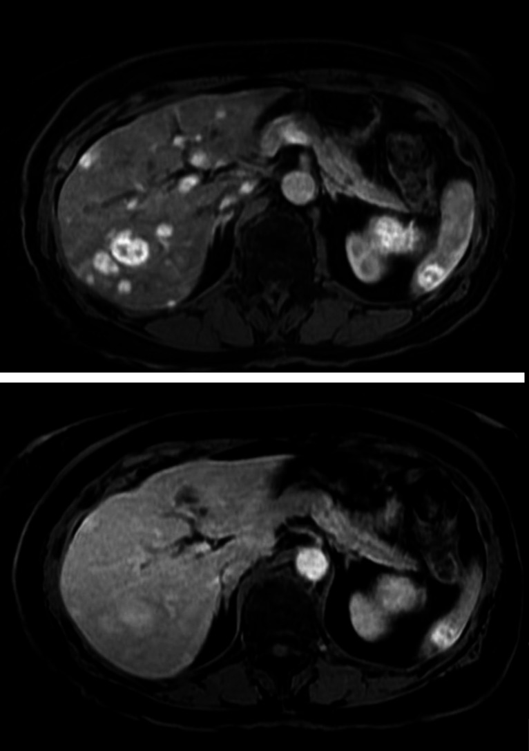
Among liver vascular tumours, hepatic small vessel neoplasm (HSVN) has been recently identified as a rare infiltrative vascular neoplasm whose malignant potential is yet to be fully ascertained. About 30 cases of HSVN have been described so far. The most common clinical presentation is an asymptomatic solitary liver lesion. Multifocal disease has been described in literature; however, to date, there are no reports of disease dissemination to other organs. Here we report a case of multifocal HSVN with synchronous spleen secondary lesions.

Keywords: Hepatic cancer; Pathology; Radiology.


Conflict of interest statement

Competing interests: None declared.

Figures

Figure 1
Figure 1
Abdomen CT scan shows multiple hepatic and splenic lesions with contrast enhancement in arterial (A) and portal phase (B) and wash-out in tardive phase (C). Radiological appearance and contrastographic behaviour are identical in both organs.
Figure 2
Figure 2
Histological specimen (H&E stained tissue) shows a vascular proliferation with anastomising vascular channels surrounded by rounded endothelial cells are observed (A). Immunohistochemistry demonstrates diffuse positivity for the vascular marker ERG (B).
Figure 3
Figure 3
H&E stained tissue. Further magnification (40x) showing bland nuclear atypias and hobnail cells surrounding the vascular channels.
Figure 4
Figure 4
Multiphasic abdomen MRI demonstrates multiple hepatic and splenic lesions with a strong and early contrast enhancement in the arterial phase (top), with a central area displaying irregular morphology and gradual enhancement; contrast enhancement persisted in the portal phase with a tendency to the wash-out in the late portal one (bottom).

References

    1. Leon M, Chavez L, Surani S. Hepatic hemangioma: what internists need to know. World J Gastroenterol 2020;26:11–20. 10.3748/wjg.v26.i1.11 - DOI - PMC - PubMed
    1. Bajenaru N, Balaban V, Săvulescu F, et al. . Hepatic hemangioma -review-. J Med Life 2015;8 Spec Issue:4–11. - PMC - PubMed
    1. Ishak KG, Sesterhenn IA, Goodman ZD, et al. . Epithelioid hemangioendothelioma of the liver: a clinicopathologic and follow-up study of 32 cases. Hum Pathol 1984;15:839–52. 10.1016/S0046-8177(84)80145-8 - DOI - PubMed
    1. Lazăr DC, Avram MF, Romoșan I, et al. . Malignant hepatic vascular tumors in adults: characteristics, diagnostic difficulties and current management. World J Clin Oncol 2019;10:110–35. 10.5306/wjco.v10.i3.110 - DOI - PMC - PubMed
    1. Gill RM, Buelow B, Mather C, et al. . Hepatic small vessel neoplasm, a rare infiltrative vascular neoplasm of uncertain malignant potential. Hum Pathol 2016;54:143–51. 10.1016/j.humpath.2016.03.018 - DOI - PMC - PubMed

Publication types