. 2022 Mar 29;12(1):5333.
doi: 10.1038/s41598-022-09299-2.

Hydrogen phosphate selectively induces MDA MB 231 triple negative breast cancer cell death in vitro

Affiliations

Hydrogen phosphate selectively induces MDA MB 231 triple negative breast cancer cell death in vitro

Aya Shanti et al. Sci Rep. .

Abstract

Phosphate ions are the most abundant anions inside the cells, and they are increasingly gaining attention as key modulators of cellular function and gene expression. However, little is known about the effect of inorganic phosphate ions on cancer cells, particularly breast cancer cells. Here, we investigated the toxicity of different phosphate compounds to triple-negative human breast cancer cells, particularly, MDA-MB-231, and compared it to that of human monocytes, THP-1. We found that, unlike dihydrogen phosphate (H2PO4-), hydrogen phosphate (HPO42-) at 20 mM or lower concentrations induced breast cancer cell death more than immune cell death, mainly via apoptosis. We correlate this effect to the fact that phosphate in the form of HPO42- raises pH levels to alkaline levels which are not optimum for transport of phosphate into cancer cells. The results in this study highlight the importance of further exploring hydrogen phosphate (HPO42-) as a potential therapeutic for the treatment of breast cancer.


Conflict of interest statement

The authors declare no competing interests.

Figures

Figure 1
Figure 1
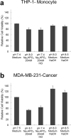
Toxicity of different phosphate compounds to (a) THP-1, human monocyte cell line, and (b) MDA-MB-231, human breast cancer cell line, evaluated after 48 h of incubation with the phosphate compound. All phosphate-containing compounds reduced viability of both cell types significantly except Na2HPO4, which only reduced the viability of MDA-MB-231 cells but not that of THP-1 cells. Experiment was done in triplicate and the sample number for each replicate is 3.
Figure 2
Figure 2
Measured pH values for different phosphate compounds prepared in cell culture media. All compounds produced acidic solutions except Na2HPO4 which produced a basic solution.
Figure 3
Figure 3
Toxicity of Sodium Phosphate Dibasic (Na2HPO4) to THP-1, human monocyte cell line and MDA-MB-231, human breast cancer cell line, evaluated via (a) MTT assay and (b) live/dead assay after 48 h of incubation with the compound. At low concentrations (< or = 20 mM), Na2HPO4 is more toxic to MDA-MB-231 cells than it is to THP-1 cells. At high concentrations (> 20 mM), Na2HPO4 is toxic to both cell types. For fluorescence images, live cells are stained in green while dead cells are stained in red. Experiments were done in triplicate and the sample number for each replicate is 3.
Figure 4
Figure 4
Mechanism of cell death induced by Sodium Phosphate Dibasic on MDA-MB-231 determined after 48 h of incubation with the phosphate compound. Live cells are shown in blue, apoptotic cells are shown in green and necrotic cells are shown in red. Sodium Phosphate Dibasic induces both necrosis and apoptosis, with apoptosis being more pronounced.
Figure 5
Figure 5
Effect of Sodium and osmotic pressure on viability of MDA-MB-231, human breast cancer cell line. In order to confirm that the observed toxicity of Na2HPO4 on MDA-MB-231 was induced by phosphate and not by sodium in NaHPO4 or by simple osmotic pressure, the toxicity of NaCl, sucrose and sodium bicarbonate on MDA-MB-231 was assessed. None of the compounds caused a significant decrease in the viability of cells. Experiment was done in triplicate and the sample number for each replicate is 3.
Figure 6
Figure 6
Measured pH values for different concentrations of (a) Sodium phosphate dibasic and (b) Sodium phosphate monobasic prepared in cell culture media. As the concentration of HPO42− increases, the pH of the solutions increases. As the concentration of H2PO4 increases, the pH of the solutions decreases.
Figure 7
Figure 7
Viability of (a) THP-1, human monocyte cell line, and (b) MDA-MB-231, human breast cancer cell line, at different pH levels evaluated after 48 h of incubation. Acidic conditions caused a significant decrease in the viability of both cell types. For basic conditions, at pH 8, the viability of THP-1 cells decreased significantly but that of MDA-MB-231 did not. At pH 9, viability of both cell types decreased significantly. Experiment was done in triplicate and the sample number for each replicate is 3.
Figure 8
Figure 8
Viability of (a) THP-1, human monocyte cell line, and (b) MDA-MB-231, breast cancer cell line, after adding Sodium Phosphate Dibasic (Na2HPO4) and adjusting pH to physiological one, evaluated after 48 h of incubation with the compound. Adjusting the pH to physiological range (7.4–7.5) after adding Na2HPO4 increased the viability of cells by 18% (approaching significance). Media only and media with NaOH are controls. Experiment was done in triplicate and the sample number for each replicate is 3.
Figure 9
Figure 9
Schematic diagram—effect of hydrogen phosphate vs. dihydrogen phosphate on the cell viability of MDA-MB-231 and THP-1 cells. Hydrogen phosphate causes significant death in MDA-MB-231 cells but not in THP-1 cells, unlike dihydrogen phosphate.

References

    1. Russo-Abrahão T, et al. Characterization of inorganic phosphate transport in the triple-negative breast cancer cell line, MDA-MB-231. PLoS ONE. 2018;13:e0191270. - PMC - PubMed
    1. Lacerda-Abreu MA, et al. H+-dependent inorganic phosphate transporter in breast cancer cells: Possible functions in the tumor microenvironment. Biochim. Biophys. Acta BBA Mol. Basis Dis. 2019;1865:2180–2188. - PubMed
    1. Camalier CE, et al. Elevated phosphate activates N-ras and promotes cell transformation and skin tumorigenesis. Cancer Prev. Res. 2010;3:359–370. - PMC - PubMed
    1. Bhutia YD, et al. SLC transporters as a novel class of tumour suppressors: Identity, function and molecular mechanisms. Biochem. J. 2016;473:1113–1124. - PMC - PubMed
    1. Sapio L, Naviglio S. Inorganic phosphate in the development and treatment of cancer: A Janus Bifrons? World J. Clin. Oncol. 2015;6:198–201. - PMC - PubMed

Publication types