. 2022 May;19(5):588-601.
doi: 10.1038/s41423-022-00855-4. Epub 2022 Mar 29.

Intranasal administration of a single dose of a candidate live attenuated vaccine derived from an NSP16-deficient SARS-CoV-2 strain confers sterilizing immunity in animals

Affiliations

Intranasal administration of a single dose of a candidate live attenuated vaccine derived from an NSP16-deficient SARS-CoV-2 strain confers sterilizing immunity in animals

Zi-Wei Ye et al. Cell Mol Immunol. 2022 May.

Abstract

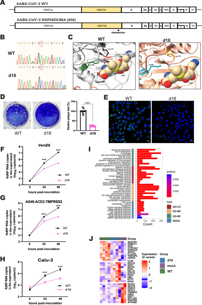
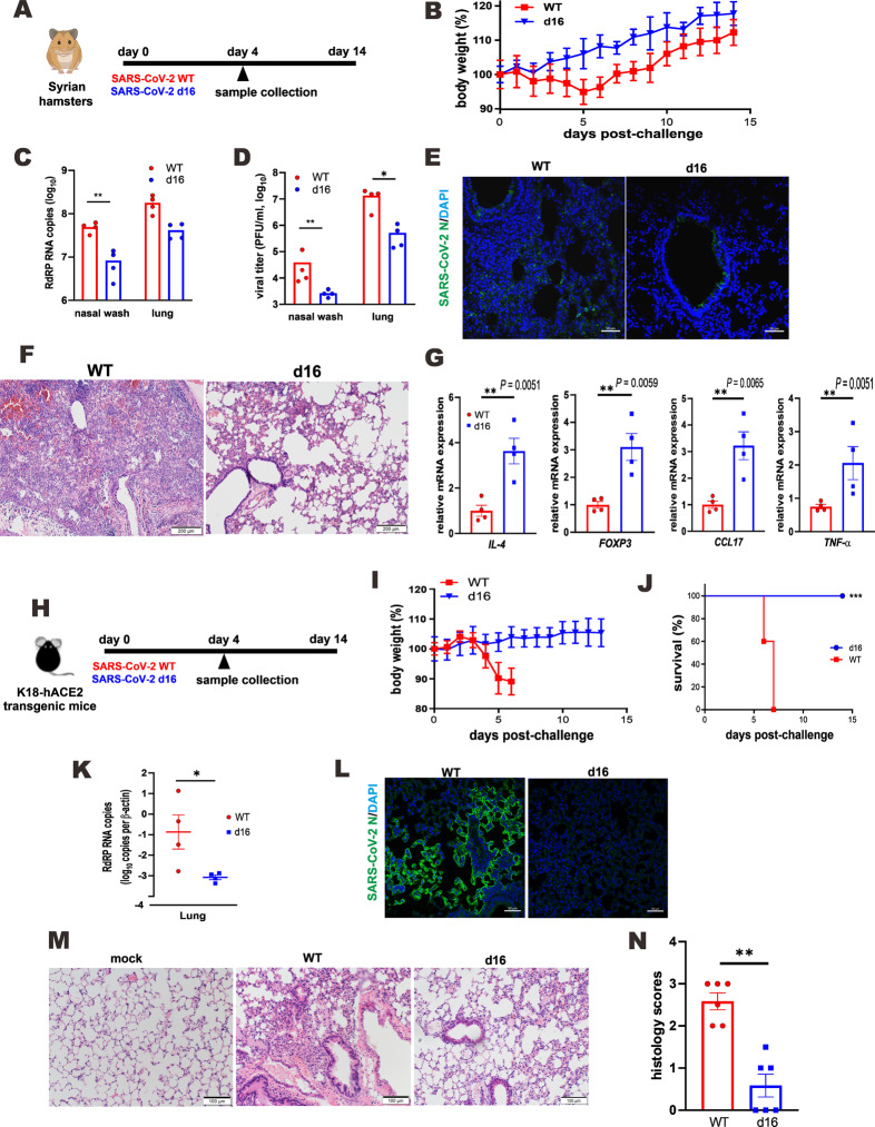
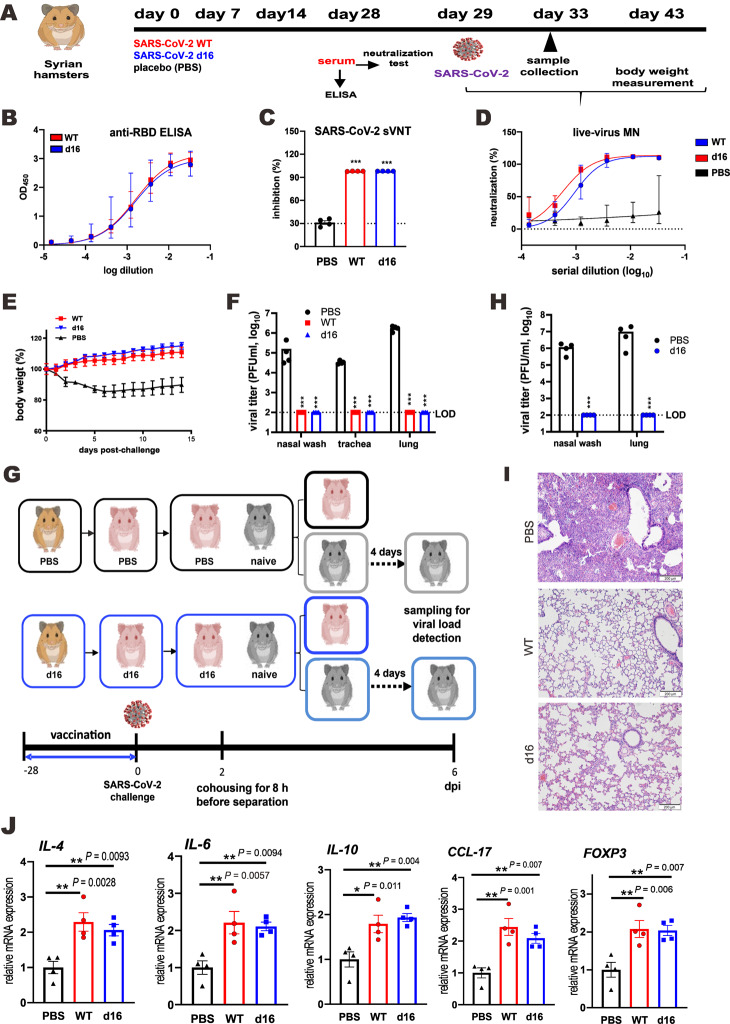
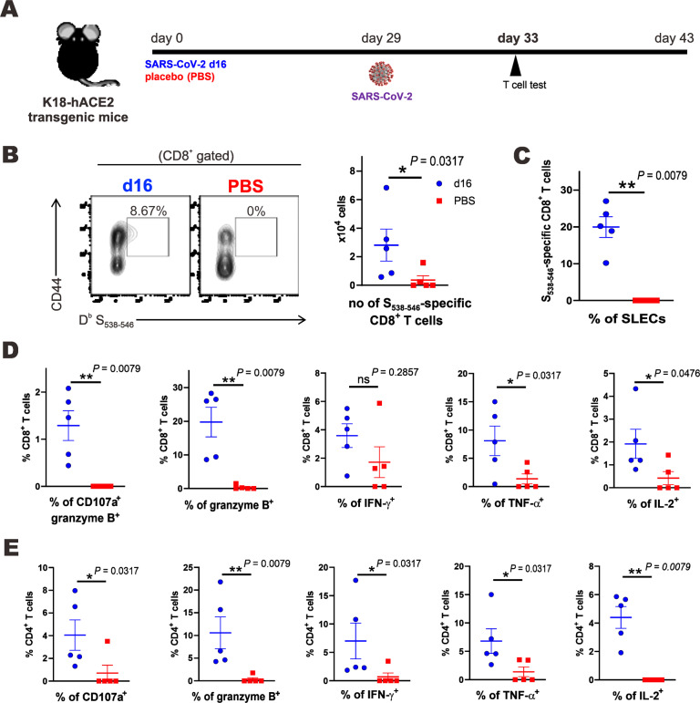
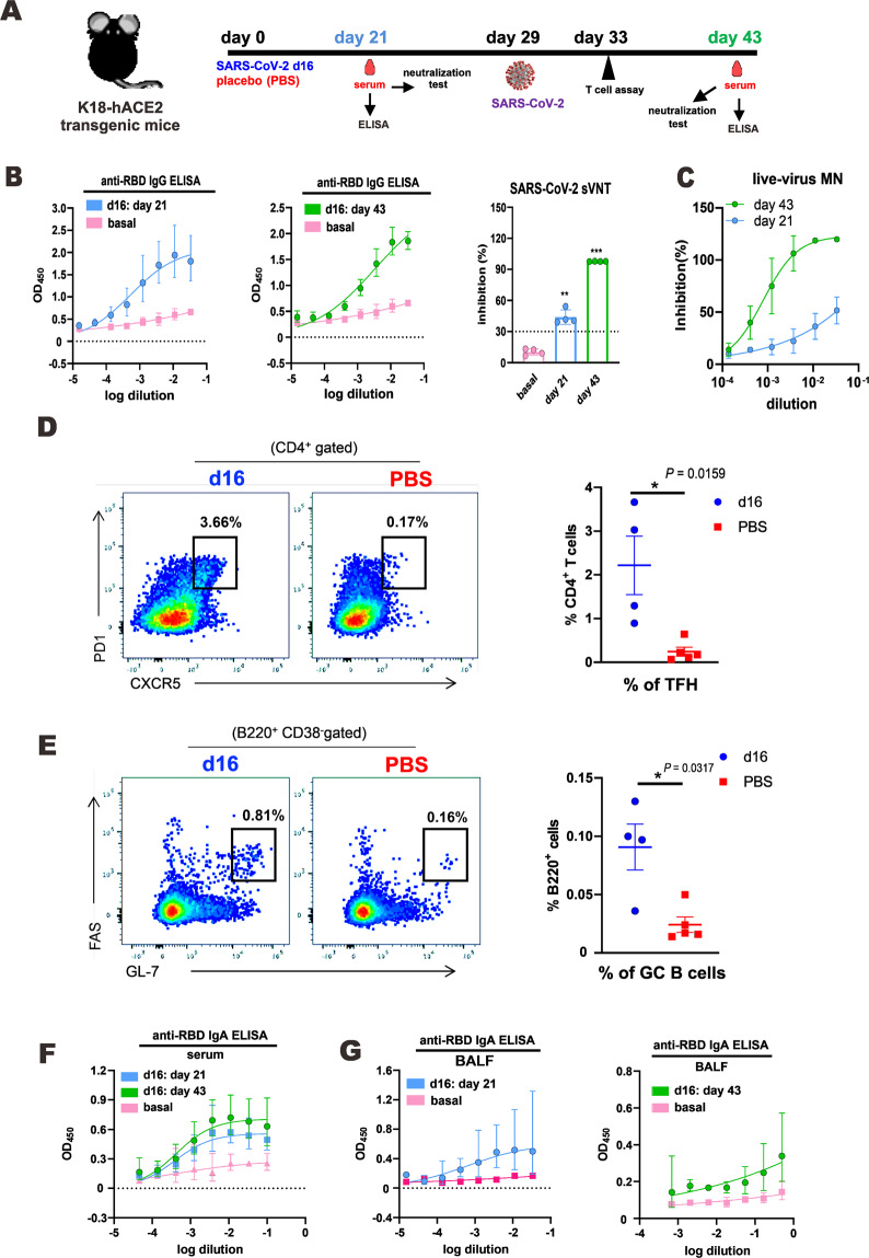
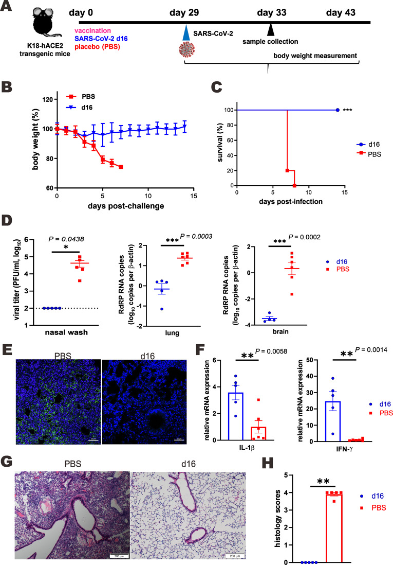
Live attenuated vaccines might elicit mucosal and sterilizing immunity against SARS-CoV-2 that the existing mRNA, adenoviral vector and inactivated vaccines fail to induce. Here, we describe a candidate live attenuated vaccine strain of SARS-CoV-2 in which the NSP16 gene, which encodes 2'-O-methyltransferase, is catalytically disrupted by a point mutation. This virus, designated d16, was severely attenuated in hamsters and transgenic mice, causing only asymptomatic and nonpathogenic infection. A single dose of d16 administered intranasally resulted in sterilizing immunity in both the upper and lower respiratory tracts of hamsters, thus preventing viral spread in a contact-based transmission model. It also robustly stimulated humoral and cell-mediated immune responses, thus conferring full protection against lethal challenge with SARS-CoV-2 in a transgenic mouse model. The neutralizing antibodies elicited by d16 effectively cross-reacted with several SARS-CoV-2 variants. Secretory immunoglobulin A was detected in the blood and nasal wash of vaccinated mice. Our work provides proof-of-principle evidence for harnessing NSP16-deficient SARS-CoV-2 for the development of live attenuated vaccines and paves the way for further preclinical studies of d16 as a prototypic vaccine strain, to which new features might be introduced to improve safety, transmissibility, immunogenicity and efficacy.

Keywords: 2′-O-methyltransferase; Live attenuated vaccine; Mucosal immunity; NSP16; Sterilizing immunity; T-cell response.


Conflict of interest statement

The authors declare no competing interests.

Figures

Fig. 1
Fig. 1
SARS-CoV-2 NSP16 D130A mutant virus (d16) is attenuated in vitro. A Schematic representation of the SARS-CoV-2 genome and the NSP16 D130A mutant. B Sequencing results for WT SARS-CoV-2 and mutant NSP16 D130A clones. The desired point mutation highlighted by the red box was verified by Sanger sequencing. C Structure of the SAM binding pocket of WT (D130) NSP16 (PDB: 6W4H) compared to that of mutant (A130) NSP16 (I-TASSER prediction). Residues in the catalytic tetrad (K46, D/A130, K170 and E203) are shown in stick representation. SAM, in surface representation, was modeled in the catalytic cleft of mutant NSP16 based on structural alignment to WT NSP16. D Plaque phenotype. VeroE6 cells were infected with the indicated recombinant viruses. After 72 h of incubation at 37 °C, the cells were fixed and stained with 1% crystal violet. Plaque images are shown. Ten plaques were randomly picked, and their sizes were quantified by ImageJ and analyzed by Student’s t-test (***P < 0.001). E Immunofluorescence staining. VeroE6 cells infected with either the WT virus or d16 at an MOI of 5 were fixed at 24 h post-infection, and the SARS-CoV-2 N antigen was detected using rabbit anti-N protein IgG and goat anti-rabbit IgG conjugated with FITC as the primary and secondary antibodies, respectively. The anti-N antisera were raised in-house and reactive to all variants tested. Cellular nuclei were stained with DAPI (blue). Scale bars, 20 μm. FH VeroE6, A549-ACE2-TMPRSS2 and Calu-3 cells were infected with recombinant WT SARS-CoV-2 (black) or d16 mutant (pink) virus at an MOI of 0.1 at the indicated time points. The cell supernatant was harvested, and SARS-CoV-2 RNA in the RNA-dependent RNA polymerase (RdRP)-coding region was quantitated using RT-qPCR and is plotted as log10 copies per ml. I Transcriptomic analysis of host gene expression in A549-ACE2-TMPRSS2 cells infected with WT SARS-CoV-2 or d16. Bar plots of enriched pathways including GO-CC, GO-MF and GO-BP terms and KEGG pathways between d16 and the WT virus are shown. GO Gene Ontology, KEGG Kyoto Encyclopedia of Genes and Genomes, BP biological process, MF molecular function, CC cellular component. J Heatmap of the enriched differentially expressed genes (false discovery rate <0.05) in the d16-, WT- or mock-infected group in comparison with the other two groups
Fig. 2
Fig. 2
SARS-CoV-2 d16 mutant virus is attenuated in vivo. A Viral challenge scheme for the hamster model. Hamsters were intranasally inoculated with 105 PFU (in 50 µl) of recombinant WT or d16 SARS-CoV-2. At 4 dpi, the lungs and nasal wash were harvested for viral yield detection. B Body weight changes of WT or d16 SARS-CoV-2-infected hamsters (n = 6). Viral loads determined assessment of the nasal wash and lung tissues of WT- or d16-challenged hamsters at 4 dpi by RT–qPCR (C) and plaque assay (D) (n = 4/group). The primers have been described elsewhere [67]. E Detection of the SARS-CoV-2 N protein in lung tissues infected with the indicated recombinant virus. The SARS-CoV-2 N protein (green) was labeled with rabbit anti-SARS-CoV-2 N antibodies followed by goat anti-rabbit antibodies conjugated to FITC. Nuclei were counterstained with DAPI (blue). Scale bars, 50 μm. F Effects of the WT virus and d16 on histopathological features of the lungs of hamsters. Representative hamster lung tissue sections harvested at 4 dpi were stained with H&E. Scale bars, 200 μm. G Proinflammatory cytokine and chemokine gene expression in WT virus- or d16-infected hamsters. The relative expression levels of representative chemokines and cytokines in lung tissue homogenates were quantified using RT–qPCR. The results are shown as the mean ± SEM. H Viral challenge scheme for the K18-hACE2 mouse model. Each mouse was intranasally inoculated with 103 PFU (in 20 µl) of recombinant WT virus or d16. At 4 dpi, the viral loads in lung tissues were quantitated using RT–qPCR. Body weight changes (I) and survival rates (J) of WT virus- or d16-infected K18-hACE2 transgenic mice (n = 5/group). K Viral loads in lung tissues at 4 dpi (n = 4/group). L Detection of the SARS-CoV-2 N protein in the lung tissues of WT virus- or d16-infected mice. The SARS-CoV-2 N protein (green) was labeled with rabbit anti-SARS-CoV-2 N antibodies followed by goat anti-rabbit antibodies conjugated to FITC. Nuclei were counterstained with DAPI (blue). Scale bars, 50 μm. M Effects of WT and d16 SARS-CoV-2 on histopathological features of mouse lungs. Lung sections from uninfected mice (mock) were included as a negative control. Representative sections of lung tissues from K18-hACE2 mice harvested at 4 dpi were stained with H&E. Scale bars, 100 μm. N Pathological changes were scored as described in the Materials and Methods. Statistical analyses by Student’s t-test (*P < 0.05; **P < 0.01; ***P < 0.001)
Fig. 3
Fig. 3
d16 as a live attenuated vaccine to alleviate disease in hamsters infected with SARS-CoV-2. A Vaccination and viral challenge scheme for the hamster model. Hamsters were intranasally inoculated with 105 PFU (in 50 µl) of recombinant WT or d16 SARS-CoV-2 on day 0. Serum samples were collected from each group on days 0, 14 and 28 for ELISA and neutralization tests. On day 29, the immunized hamsters were challenged with 105 PFU (in 50 µl) of clinical isolate HK-13 of SARS-CoV-2. Respiratory tissues (tracheae and lungs) and the nasal wash were collected on day 33, and each respective viral load was detected by a plaque assay. Body weights were recorded daily from day 29 to day 43 after viral challenge. B Antibody responses in hamsters infected with WT SARS-CoV-2 or immunized with d16 SARS-CoV-2 were measured by ELISA to assess RBD-specific antibodies. C The cross-neutralization capability of infected/immunized hamster sera (1:10 dilution) was examined using the SARS-CoV-2 sVNT assay. The dotted line represents the assay cutoff at 30% inhibition. D SARS-CoV-2 neutralization tests to examine the neutralization ability of hamster sera collected from different groups. E Body weight changes of HK-13-infected hamsters (n = 5) vaccinated with d16 viruses or PBS. The WT virus-reinfected group served as a control. F Viral titer determination by a plaque assay for the nasal wash, tracheal tissues and lung tissues of HK-13-challenged hamsters in the WT, d16 or PBS group at 4 dpi (n = 4/group). LOD limit of detection. G Hamster cohousing scheme. Briefly, d16- or sham-vaccinated hamsters (n = 4/group) were infected with SARS-CoV-2 on day 29 post-vaccination and subsequently cohoused with immunologically naïve hamsters for 8 h at 2 dpi, followed by separation and viral load analysis of the recipient hamsters on day 4 post-cohousing. H Viral loads in the nasal wash and lung tissues of recipient hamsters at 4 days post-cohousing. LOD limit of detection. I Histopathological changes in the lungs of SARS-CoV-2-challenged hamsters in the WT, d16 or PBS group at 4 dpi. Representative sections were stained with H&E. Scale bars, 200 μm. J Transcript levels of representative proinflammatory chemokines and cytokines in lung tissue homogenates from the WT, d16 and PBS groups, as measured by RT–qPCR. The results are shown in arbitrary units as the mean ± SEM. Statistical analyses were performed with Student’s t-test (*P < 0.05; **P < 0.01; ***P < 0.001).
Fig. 4
Fig. 4
T-cell immunity elicited by vaccination with d16 in the K18-hACE2 transgenic mouse model. A Vaccination and viral challenge scheme for d16-vaccinated K18-hACE2 mice assessed with a T-cell assay. K18-hACE2 mice were intranasally vaccinated with 103 PFU (in 20 µl) of d16 or PBS (n = 5 mice/group) and challenged with 104 PFU of clinical isolate HK-13 of SARS-CoV-2 on day 29. Lung-origin T cells were subjected to flow cytometric analysis at 4 days post-infection. B Representative flow tracings and the absolute number of S538–546-specific CD8+ T cells. C Proportions of short-lived effector cells (SLECs, KLRG1+ IL-7R-) among S538–546-specific CD8+ T cells. Lung-origin lymphocytes were stimulated with 1 μg/ml S538–546 (D) or S62–76 (E) peptides for 4 h in the presence of brefeldin A. The percentages of CD107a+ and cytokine-producing CD8+ (D) and CD4+ (E) T cells were also assessed. The results are shown as the mean ± SEM. Statistical analyses were performed with Student’s t-test (*P < 0.05; **P < 0.01; ***P < 0.001)
Fig. 5
Fig. 5
Humoral and cell-mediated immune responses in d16-vaccinated mice. A Vaccination and viral challenge scheme for d16-vaccinated or PBS-treated K18-hACE2 mice assessed by antibody analysis. Briefly, K18-hACE2 mice intranasally inoculated with 103 PFU (in 20 µl) of d16 or PBS (n = 5 mice/group) were challenged with 104 PFU of clinical isolate HK-13 of SARS-CoV-2 on day 29. Serum samples were collected from each group on days 21 and 43 for ELISA and neutralization analysis. B Anti-RBD IgG ELISA and sVNT assays for neutralizing activity on day 21 post-vaccination (blue) and 14 days after challenge (green). The dotted line represents the assay cutoff at 30% inhibition for the sVNT assay. Basal levels were obtained for mice before the start of the experiment. C The cross-neutralization ability of d16-immunized mouse serum (1:10 dilution) was examined using a live-virus MN assay for the HK-13 clinical isolate of SARS-CoV-2. D Representative flow charts and percentages of T follicular helper cells (TFH cells, CD4+ PD1+ CXCR5+) in the spleen. E Representative flow charts and percentages of germinal center B cells (GC B cells, B220+ CD38- GL7+ FAS+) in the spleen. Results are shown as the mean ± SEM. Anti-RBD IgA in mouse serum (F) and bronchoalveolar lavage fluid (BALF) (G) collected on day 21 post-vaccination (blue) and 14 days after the challenge (green). Basal levels were obtained for mice before the start of the experiments. Statistical analyses performed with one-way ANOVA with Dunnett’s post-hoc test or Student’s t-test (*P < 0.05; **P < 0.01; ***P < 0.001)
Fig. 6
Fig. 6
d16 confers full protection against SARS-CoV-2 lethal challenge in the K18-hACE2 transgenic mouse model. A Vaccination and viral challenge scheme for K18-hACE2 mice (n ≥ 10). After intranasal vaccination with 103 PFU (in 20 µl) of recombinant d16 SARS-CoV-2 or PBS, each vaccinated mouse was challenged with 104 PFU (in 20 µl) of clinical isolate HK-13 of SARS-CoV-2 on day 29. Body weights were recorded daily for 14 days after the viral challenge. On day 33, the nasal wash, lung tissues and brain tissues were collected for quantification of viral loads using RT–qPCR. Body weight changes (B) and survival rates (C) of SARS-CoV-2-infected mice vaccinated with d16 or PBS (n = 5/group). D Viral loads measured by plaque assays or RT–qPCR for the nasal wash, lung tissues and brain tissues collected from SARS-CoV-2-infected mice vaccinated with d16 or PBS at 4 dpi (n ≥ 5/group). Statistical analyses were performed with Student’s t-test (*P < 0.05; ***P < 0.001). E Detection of the SARS-CoV-2 N protein in the lung tissues of SARS-CoV-2-infected mice vaccinated with d16 or PBS. The SARS-CoV-2 N protein (green) was labeled with rabbit anti-SARS-CoV-2 N antibodies followed by goat anti-rabbit antibodies conjugated to FITC. Nuclei were counterstained with DAPI (blue). Scale bars, 50 μm. F IL-1β and IFN-γ gene expression in SARS-CoV-2-infected mouse lungs. The transcripts of representative chemokines and cytokines in lung tissue homogenates from the indicated groups were quantitated using RT–qPCR. The results are shown as the mean ± SEM. Statistical analyses were performed with Student’s t-test (*P < 0.05; **P < 0.01; ***P < 0.001). G Effect of vaccination on the histopathological changes in the lungs of SARS-CoV-2-infected mice. Representative sections of lung tissue from mice harvested at 4 dpi were stained with H&E. Scale bars, 100 μm. H Pathological changes were scored as described in the Materials and Methods (**P < 0.01)

Similar articles

Cited by

References

    1. Brown CM, Vostok J, Johnson H, Burns M, Gharpure R, Sami S, et al. Outbreak of SARS-CoV-2 infections, including COVID-19 vaccine breakthrough infections, associated with large public gatherings – Barnstable County, Massachusetts, July 2021. MMWR Morb Mortal Wkly Rep. 2012;70:1059–62. doi: 10.15585/mmwr.mm7031e2. - DOI - PMC - PubMed
    1. Mishra S, Sören M, Sharma M, Whittaker C, Mellan TA, Wilton T, et al. Changing composition of SARS-CoV-2 lineages and rise of delta variant in England. EClinicalMedicine. 2021;39:101064. doi: 10.1016/j.eclinm.2021.101064. - DOI - PMC - PubMed
    1. Nanduri S, Pilishvili T, Derado G, Soe MM, Dollard P, Wu H, et al. Effectiveness of Pfizer-BioNTech and Moderna vaccines in preventing SARS-CoV-2 infection among nursing home residents before and during widespread circulation of the SARS-CoV-2 B.1.617.2 (δ) variant – National Healthcare Safety Network, March 1-August 1, 2021. MMWR Morb Mortal Wkly Rep. 2021;70:1163–6. doi: 10.15585/mmwr.mm7034e3. - DOI - PMC - PubMed
    1. Kuhlmann C, Mayer CK, Claassen M, Maponga T, Burgers WA, Keeton R, et al. Breakthrough infections with SARS-CoV-2 Omicron despite mRNA vaccine booster dose. Lancet. 2022;399:625–6. 10.1016/S0140-6736(22)00090-3 - PMC - PubMed
    1. Tré-Hardy M, Cupaiolo R, Wilmet A, Antoine-Moussiaux T, Della Vecchia A, Horeanga A, et al. Six-month interim analysis of ongoing immunogenicity surveillance of the mRNA-1273 vaccine in healthcare workers: a third dose is expected. J Infect. 2021;S0163-4453:00433–3. doi: 10.1016/j.jinf.2021.08.031. - DOI - PubMed

Publication types

Supplementary concepts