Review
. 2022 May;18(5):273-288.
doi: 10.1038/s41582-022-00635-8. Epub 2022 Mar 29.

Post-traumatic stress disorder: clinical and translational neuroscience from cells to circuits

Affiliations
Review

Post-traumatic stress disorder: clinical and translational neuroscience from cells to circuits

Kerry J Ressler et al. Nat Rev Neurol. 2022 May.

Abstract

Post-traumatic stress disorder (PTSD) is a maladaptive and debilitating psychiatric disorder, characterized by re-experiencing, avoidance, negative emotions and thoughts, and hyperarousal in the months and years following exposure to severe trauma. PTSD has a prevalence of approximately 6-8% in the general population, although this can increase to 25% among groups who have experienced severe psychological trauma, such as combat veterans, refugees and victims of assault. The risk of developing PTSD in the aftermath of severe trauma is determined by multiple factors, including genetics - at least 30-40% of the risk of PTSD is heritable - and past history, for example, prior adult and childhood trauma. Many of the primary symptoms of PTSD, including hyperarousal and sleep dysregulation, are increasingly understood through translational neuroscience. In addition, a large amount of evidence suggests that PTSD can be viewed, at least in part, as a disorder that involves dysregulation of normal fear processes. The neural circuitry underlying fear and threat-related behaviour and learning in mammals, including the amygdala-hippocampus-medial prefrontal cortex circuit, is among the most well-understood in behavioural neuroscience. Furthermore, the study of threat-responding and its underlying circuitry has led to rapid progress in understanding learning and memory processes. By combining molecular-genetic approaches with a translational, mechanistic knowledge of fear circuitry, transformational advances in the conceptual framework, diagnosis and treatment of PTSD are possible. In this Review, we describe the clinical features and current treatments for PTSD, examine the neurobiology of symptom domains, highlight genomic advances and discuss translational approaches to understanding mechanisms and identifying new treatments and interventions for this devastating syndrome.


Conflict of interest statement

Competing interests

K.J.R. has received consulting income from Alkermes, Bionomics, Bioxcel and Jazz Pharmaceuticals, and is on scientific advisory boards for the Army STARRS Project, Janssen, the National Center for PTSD, Sage Therapeutics and Verily. He has also received sponsored research support from Brainsway and Takeda. He also serves on the Boards of ACNP and Biological Psychiatry. W.C. has received consulting income from Psy Therapeutics and has a sponsored research agreement with Cerevel Therapeutics. He is the editor-in-chief for Neuropsychopharmacology and serves on the board of ACNP. None of this work is directly related to the work presented here. S.L.R. receives compensation as a Board member of Community Psychiatry and for his role as Secretary of SOBP. He also serves on the Boards of ADAA and NNDC. He has received royalties from Oxford University Press and APPI.

Figures
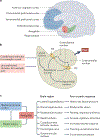
Fig. 1 |
Fig. 1 |. Schematic diagram of neural circuitry involved in fear conditioning and post-traumatic stress disorder.
a | The primary brain regions involved in regulating the fear response are the amygdala (red), hippocampus (green) and medial prefrontal cortex (blue), which comprises the dorsal and ventral subdivisions, orbitofrontal cortex, and anterior cingulate cortex. b | The amygdala sits at the centre of the neural circuit involved in regulating fear conditioning. Generally speaking, inputs into basolateral nuclei of the amygdala lead to learning about fear, whereas the central amygdala is responsible for sending output signals about fear, including to the hypothalamus and brainstem structures. c | The interactions of the medial prefrontal cortex component parts and the hippocampus constantly regulate amygdala output to subcortical brain regions that activate the fear reflex. The medial prefrontal cortex (in particular the ventromedial prefrontal cortex) is classically thought to inhibit amygdala activity (and reduce subjective distress), whereas the hippocampus has a role both in the coding of fear memories as well as in the regulation of the amygdala. ITC, intercalated cells.
Fig. 2 |
Fig. 2 |. Neurophysiological findings commonly seen in individuals with PTSD.
a | Functional MRI is commonly used to quantify and visualize brain activation to emotionally relevant stimuli. In this example, individuals with post-traumatic stress disorder (PTSD) (right) have significantly more amygdala activation (orange pixels) in response to the image of a fearful face than do control participants who have experienced trauma but do not have PTSD (left). b | Another commonly used method for examining physiological responses in anxiety and trauma-related disorders is the fear-potentiated startle response. To detect this response, electrodes are placed under the eye of a participant (left) to measure the muscle activity during an eyeblink. The example on the right shows the response of a healthy individual at baseline (black) and in a threat state (red). Individuals with PTSD have an enhanced response under both conditions. c | A structural MRI scan shows smaller hippocampal volume in a participant with PTSD (right) than in a participant without PTSD (left). The hippocampus is outlined in red. d | Compared with control participants, individuals with PTSD have lower regional cerebral blood flow activity in the rostral anterior cingulate during exposure to traumatic or stressful script-driven imagery. Yellow pixels show change from the control group. e | Blood-based biomarkers, such as plasma cortisol, serve as a measure of the hypothalamic–pituitary–adrenal axis response to stress. Individuals with PTSD have a hyper-sensitive hypothalamic–pituitary–adrenal axis response to the cortisol agonist dexamethasone, such that following a dose of dexamethasone, they show a ‘super-suppression’ of plasma cortisol levels (blue) compared with healthy individuals (green). Part c adapted with permission from REF., Elsevier. Part d reprinted with permission from REF., Elsevier.
Fig. 3 |
Fig. 3 |. Schematic diagram of circuits and neurotransmitters regulating fear and threat responses.
Within the amygdala, parallel neural circuits enhance (Fear-On; red) or block (Fear-off; green) fear responses or can be involved in appetitive responses (APPT-On; blue). Neuronal populations involved in these circuits have been found to have distinct molecular signatures. Specifically, interconnected neuronal populations involved in Fear-On circuits express factors such as corticotropin-releasing factor (CRF; also known as CRH), pituitary adenylate cyclase-activating polypeptide (PACAP) and TAC2, known to have key roles in responses to fear, stress and anxiety. Fear-On, Fear-Off and APPT-On amygdala circuits are reciprocally regulated and are part of larger neural networks with corresponding functions. A disruption of these circuits, with enhancement of Fear-On circuits and dampening of Fear-Off circuits, could underlie many of the anxiety-related symptoms in post-traumatic stress disorder (PTSD). A dysregulation of the APPT-On circuits might result in symptoms related to depression, particularly anhedonia and avolition, often comorbid with PTSD. A, anterior; ITC, intercalated cells; P, posterior.

Similar articles

Cited by

References

    1. American Psychiatric Association. Diagnostic and Statistical Manual of Mental Disorders 5th edn (American Psychiatric Publishing, 2013).
    1. Breslau N et al. Trauma and posttraumatic stress disorder in the community: the 1996 Detroit area survey of trauma. Arch. Gen. Psychiatry 55, 626–632 (1998). - PubMed
    1. Breslau N, Peterson EL, Poisson LM, Schultz LR & Lucia VC Estimating post-traumatic stress disorder in the community: lifetime perspective and the impact of typical traumatic events. Psychol. Med 34, 889–898 (2004). - PubMed
    1. Bromet E, Sonnega A & Kessler RC Risk factors for DSM-III-R posttraumatic stress disorder: findings from the National Comorbidity Survey. Am. J. Epidemiol. 147, 353–361 (1998). - PubMed
    1. McLaughlin KA et al. Subthreshold posttraumatic stress disorder in the world health organization world mental health surveys. Biol. Psychiatry 77, 375–384 (2015). - PMC - PubMed

Publication types