

Association of Brain Volumes and White Matter Injury With Race, Ethnicity, and Cardiovascular Risk Factors: The Multi-Ethnic Study of Atherosclerosis
- PMID: 35352569
- PMCID: PMC9075451
- DOI: 10.1161/JAHA.121.023159
Association of Brain Volumes and White Matter Injury With Race, Ethnicity, and Cardiovascular Risk Factors: The Multi-Ethnic Study of Atherosclerosis
Abstract
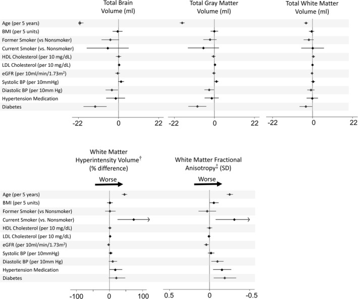
Background Cardiovascular risk factors are associated with cognitive decline and dementia. Magnetic resonance imaging provides sensitive measurement of brain morphology and vascular brain injury. However, associations of risk factors with brain magnetic resonance imaging findings have largely been studied in White participants. We investigated associations of race, ethnicity, and cardiovascular risk factors with brain morphology and white matter (WM) injury in a diverse population. Methods and Results In the Multi-Ethnic Study of Atherosclerosis, measures were made in 2018 to 2019 of total brain volume, gray matter and WM volume, and WM injury, including WM hyperintensity volume and WM fractional anisotropy. We assessed cross-sectional associations of race and ethnicity and of cardiovascular risk factors with magnetic resonance imaging measures. Magnetic resonance imaging data were complete in 1036 participants; 25% Black, 15% Chinese-American, 19% Hispanic, and 41% White. Mean (SD) age was 72 (8) years and 53% were women. Although WM injury was greater in Black than in White participants in a minimally adjusted model, additional adjustment for cardiovascular risk factors and socioeconomic status each attenuated this association, rendering it nonsignificant. Overall, greater average WM hyperintensity volume was associated with older age and current smoking (69% greater vs never smoking); lower fractional anisotropy was additionally associated with higher diastolic blood pressure, use of antihypertensive medication, and diabetes. Conclusions We found no statistically significant difference in measures of WM injury by race and ethnicity after adjustment for cardiovascular risk factors and socioeconomic status. In all racial and ethnic groups, older age, current smoking, hypertension, and diabetes were strongly associated with WM injury.
Keywords: brain magnetic resonance imaging; cardiovascular risk factors; race and ethnicity; white matter injury.
Figures

References
-
- Smith EE, Egorova S, Blacker D, Killiany RJ, Muzikansky A, Dickerson BC, Tanzi RE, Albert MS, Greenberg SM, Guttmann CR. Magnetic resonance imaging white matter hyperintensities and brain volume in the prediction of mild cognitive impairment and dementia. Arch Neurol. 2008;65:94–100. doi: 10.1001/archneurol.2007.23 - DOI - PubMed
-
- Wu A, Sharrett AR, Gottesman RF, Power MC, Mosley TH Jr, Jack CR Jr, Knopman DS, Windham BG, Gross AL, Coresh J. Association of brain magnetic resonance imaging signs with cognitive outcomes in persons with nonimpaired cognition and mild cognitive impairment. JAMA Netw Open. 2019;2:e193359. doi: 10.1001/jamanetworkopen.2019.3359 - DOI - PMC - PubMed
-
- Brickman AM, Honig LS, Scarmeas N, Tatarina O, Sanders L, Albert MS, Brandt J, Blacker D, Stern Y. Measuring cerebral atrophy and white matter hyperintensity burden to predict the rate of cognitive decline in Alzheimer disease. Arch Neurol. 2008;65:1202–1208. doi: 10.1001/archneur.65.9.1202 - DOI - PMC - PubMed
-
- Maillard P, Seshadri S, Beiser A, Himali JJ, Au R, Fletcher E, Carmichael O, Wolf PA, DeCarli C. Effects of systolic blood pressure on white‐matter integrity in young adults in the Framingham Heart Study: a cross‐sectional study. Lancet Neurol. 2012;11:1039–1047. doi: 10.1016/S1474-4422(12)70241-7 - DOI - PMC - PubMed
Publication types
MeSH terms
Grants and funding
LinkOut - more resources
Full Text Sources
Medical
