Review
. 2022 Feb;80(2):180-191.
doi: 10.1590/0004-282X-ANP-2021-0134.

Optical coherence tomography in neurodegenerative disorders

Affiliations
Review

Optical coherence tomography in neurodegenerative disorders

Leonardo Provetti Cunha et al. Arq Neuropsiquiatr. 2022 Feb.

Abstract

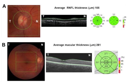
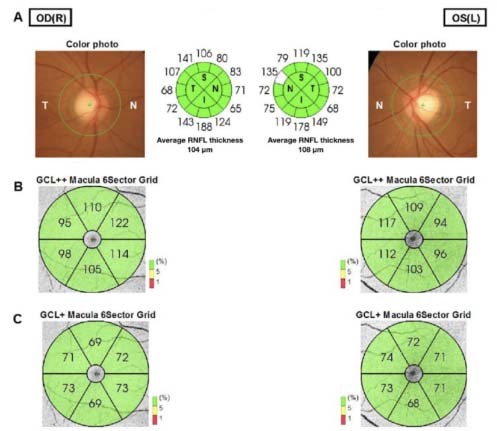
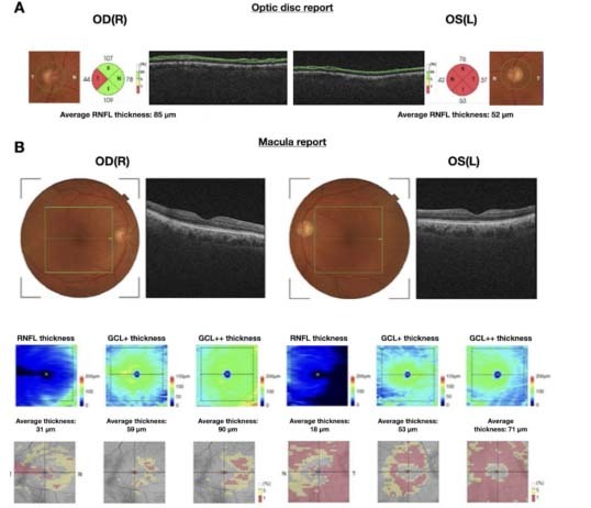
Structural imaging of the brain is the most widely used diagnostic tool for investigating neurodegenerative diseases. More advanced structural imaging techniques have been applied to early or prodromic phases, but they are expensive and not widely available. Therefore, it is highly desirable to search for noninvasive, easily accessible, low-cost clinical biomarkers suitable for large-scale population screening, in order to focus on making diagnoses at the earliest stages of the disease. In this scenario, imaging studies focusing on the structures of the retina have increasingly been used for evaluating neurodegenerative diseases. The retina shares embryological, histological, biochemical, microvascular and neurotransmitter similarities with the cerebral cortex, thus making it a uniquely promising biomarker for neurodegenerative diseases. Optical coherence tomography is a modern noninvasive imaging technique that provides high-resolution two-dimensional cross-sectional images and quantitative reproducible three-dimensional volumetric measurements of the optic nerve head and retina. This technology is widely used in ophthalmology practice for diagnosing and following up several eye diseases, such as glaucoma, diabetic retinopathy and age-related macular degeneration. Its clinical impact on neurodegenerative diseases has raised enormous interest over recent years, as several clinical studies have demonstrated that these diseases give rise to reduced thickness of the inner retinal nerve fiber layer, mainly composed of retinal ganglion cells and their axons. In this review, we aimed to address the clinical utility of optical coherence tomography for diagnosing and evaluating different neurodegenerative diseases, to show the potential of this noninvasive and easily accessible method.

A avaliação estrutural do cérebro, feita por meio dos exames de neuroimagem, é a forma mais utilizada de ferramenta diagnóstica e de acompanhamento das doenças neurodegenerativas. Técnicas de imagem mais sofisticadas podem ser necessárias especialmente nas fases mais precoces, antes mesmo do surgimento de quaisquer sintomas, porém costumam ser caras e pouco acessíveis. Sendo assim, é de fundamental importância a busca de biomarcadores não invasivos, de fácil acesso e baixo custo, que possam ser utilizados para rastreio populacional e diagnóstico mais precoce. Nesse cenário, o número de estudos com ênfase em técnicas de imagem para avaliação estrutural da retina em pacientes com doenças neurodegenerativas tem aumentado nos últimos anos. A retina apresenta similaridade embriológica, histológica, bioquímica, microvascular e neurotransmissora com o córtex cerebral, tornando-se assim um biomarcador único e promissor nas doenças neurodegenerativas. A tomografia de coerência óptica é uma moderna técnica de imagem não invasiva que gera imagens seccionais bidimensionais de alta resolução e medidas volumétricas tridimensionais reprodutivas do disco óptico e da mácula. Essa tecnologia é amplamente utilizada na prática oftalmológica para o diagnóstico e o seguimento de diversas doenças oculares, como glaucoma, retinopatia diabética e degeneração macular relacionada à idade. A redução da espessura da camada de fibras nervosas da retina e das camadas de células ganglionares em pacientes com doenças neurodegenerativas foi demonstrada em diversos estudos clínicos nos últimos anos. Nesta revisão, abordamos as principais aplicações clínicas da tomografia de coerência óptica nas doenças neurodegenerativas e discutimos o seu papel como potencial biomarcador nessas afecções.


Conflict of interest statement

Conflicts of interest: There is no conflict of interest to declare.

Figures

Figure 1.
Figure 1.. Schematic representation of the optic nerve head (ONH) and macula using spectral-domain optical coherence tomography (OCT) scanning protocols on the right eye of a normal individual. (A) 3D optic disc report, showing the ONH. Left panel: 6 × 6 mm scanned area centered on the ONH (green square) and the 3.4 mm peripapillary analyzed area for assessment of RNFL thickness. Central panel: OCT B-scan representing the cross-sectional retinal image around the ONH. The boundaries of the RNFL are represented by green lines. Right panel: Schematic representation of the peripapillary RNFL thickness measurements divided into 4 and 12-clock hour sectors with the values in microns. (B) 3D macula report, showing the macular area. Left panel: 6 × 6 mm scanned area centered on the fovea (green square). The horizontal green line represents the scanned horizontal central area. Central panel: Horizontal OCT B-scan representing the cross-sectional retinal image. The boundaries of the internal limiting membrane and the retinal pigment epithelium are represented by green lines. Right panel: ETDRS map divided in 9 sectors with the respective values of the total macular thickness measurements in microns.
Figure 2.
Figure 2.. Swept-source optical coherence tomography (OCT) 3D wide disc and macula report (12 × 9 mm), on OCT three-dimensional images of both eyes (OU) of a 45-year-old healthy woman. (A) Peripapillary retinal nerve fiber (pRNFL) layer thickness measurements of OU. Average pRNFL thickness measurements are within normal limits: 104 microns in right eye (OD) and 108 microns in left eye (OS). (B) Ganglion cell complex (GCL++) macula thickness map divided into six sectors. GCL++ thickness measurements are within normal limits (in green) in OU. (C) Ganglion cell/inner plexiform layer (GCL+) macula thickness map divided into 6 sectors. GCL+ thickness measurements are within normal limits (in green) in OU. In this example, OCT thickness measurements did not demonstrate any signs of axonal loss and neuronal loss.
Figure 3.
Figure 3.. Example of optical coherence tomography (OCT) 3D optic disc and macula report, on OCT three-dimensional images of both eyes (OU) of a 75-year-old male with a three-year diagnosis of Alzheimer’s disease and Mini-Mental State Examination test score of 15/30. (A) Optic disc report (6 × 6 mm) of OU, including total average peripapillary retinal nerve fiber layer (RNFL) measurements (in microns). Note the values outside of normal limits in the temporal quadrant in the right eye and in all four quadrants in the left eye (in red). (B) Inner macular thickness report (7 × 7 mm) of macular RNFL, ganglion cell/plexiform layer (GCL+) and ganglion cell/plexiform layer plus RNFL (GCL++) thickness measurements (in microns). Note the values outside of normal limits (in red) for all three parameters in OU, thus showing greater severity of neuronal loss in the left eye.

References

    1. Heemels MT. Neurodegenerative diseases. Nature. 2016 Nov;539(7628):179–179. doi: 10.1038/539179a. - DOI - PubMed
    1. Vadakkan KI. Neurodegenerative disorders share common features of “loss of function” states of a proposed mechanism of nervous system functions. Biomed Pharmacother. 2016 Oct;83:412–430. doi: 10.1016/j.biopha.2016.06.042. - DOI - PubMed
    1. Risacher SL, Saykin AJ. Neuroimaging and other biomarkers for Alzheimer’s disease: the changing landscape of early detection. Annu Rev Clin Psychol. 2013 Jan;9:621–648. doi: 10.1146/annurev-clinpsy-050212-185535. - DOI - PMC - PubMed
    1. Alber J, Goldfarb D, Thompson LI, Arthur E, Hernandez K, Cheng D, et al. Developing retinal biomarkers for the earliest stages of Alzheimer’s disease: what we know, what we don’t, and how to move forward. Alzheimers Dement. 2020 Jan;16(1):229–243. doi: 10.1002/alz.12006. - DOI - PubMed
    1. Huang D, Swanson EA, Lin CP, Schuman JS, Stinson WG, Chang W, et al. Optical coherence tomography. Science. 1991 Nov;254(5035):1178–1181. doi: 10.1126/science.1957169. - DOI - PMC - PubMed

MeSH terms