. 2022 May 1;7(5):494-503.
doi: 10.1001/jamacardio.2022.0183.

Assessment of Artificial Intelligence in Echocardiography Diagnostics in Differentiating Takotsubo Syndrome From Myocardial Infarction

Affiliations

Assessment of Artificial Intelligence in Echocardiography Diagnostics in Differentiating Takotsubo Syndrome From Myocardial Infarction

Fabian Laumer et al. JAMA Cardiol. .

Abstract

Importance: Machine learning algorithms enable the automatic classification of cardiovascular diseases based on raw cardiac ultrasound imaging data. However, the utility of machine learning in distinguishing between takotsubo syndrome (TTS) and acute myocardial infarction (AMI) has not been studied.

Objectives: To assess the utility of machine learning systems for automatic discrimination of TTS and AMI.

Design, settings, and participants: This cohort study included clinical data and transthoracic echocardiogram results of patients with AMI from the Zurich Acute Coronary Syndrome Registry and patients with TTS obtained from 7 cardiovascular centers in the International Takotsubo Registry. Data from the validation cohort were obtained from April 2011 to February 2017. Data from the training cohort were obtained from March 2017 to May 2019. Data were analyzed from September 2019 to June 2021.

Exposure: Transthoracic echocardiograms of 224 patients with TTS and 224 patients with AMI were analyzed.

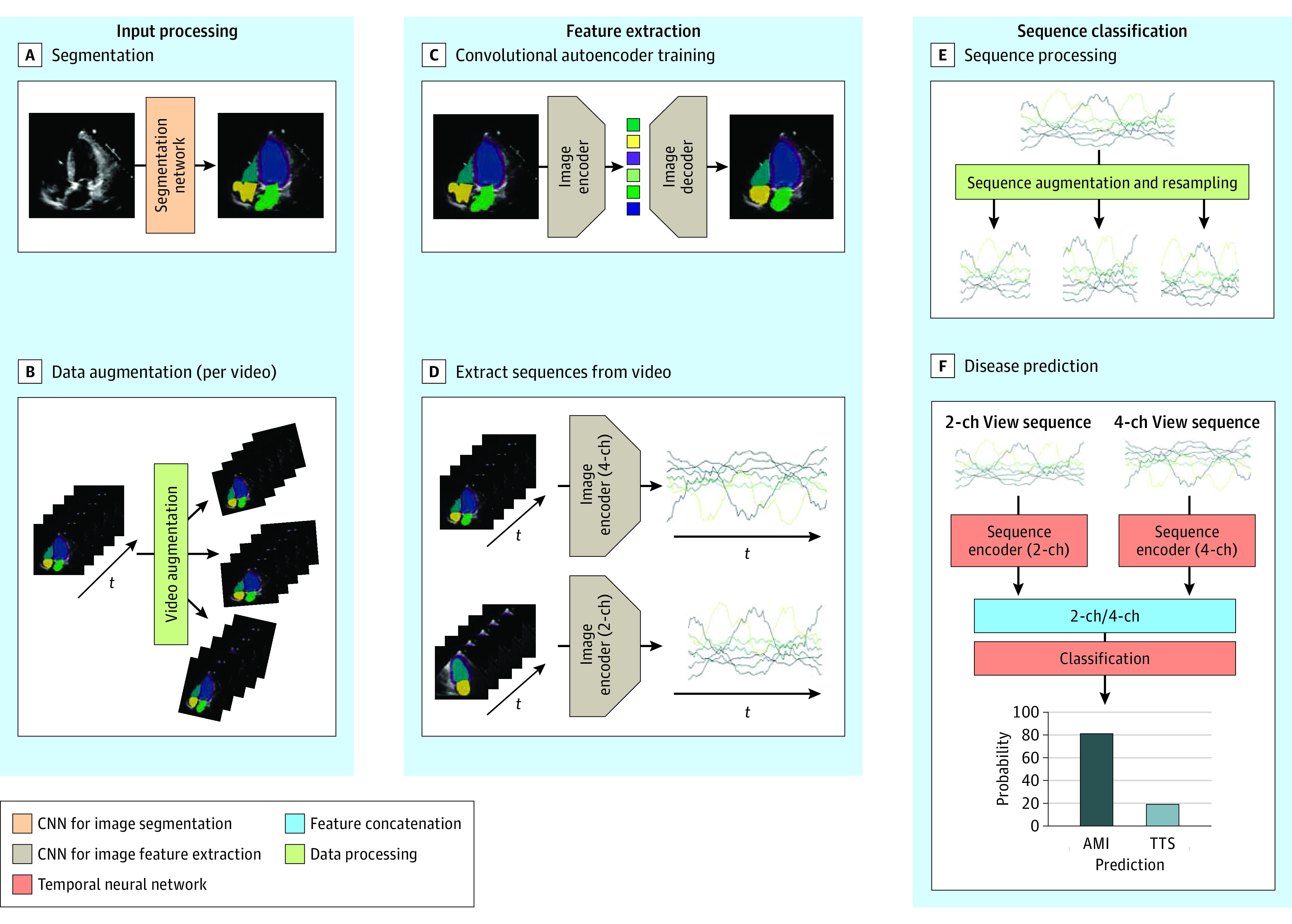
Main outcomes and measures: Area under the receiver operating characteristic curve (AUC), accuracy, sensitivity, and specificity of the machine learning system evaluated on an independent data set and 4 practicing cardiologists for comparison. Echocardiography videos of 228 patients were used in the development and training of a deep learning model. The performance of the automated echocardiogram video analysis method was evaluated on an independent data set consisting of 220 patients. Data were matched according to age, sex, and ST-segment elevation/non-ST-segment elevation (1 patient with AMI for each patient with TTS). Predictions were compared with echocardiographic-based interpretations from 4 practicing cardiologists in terms of sensitivity, specificity, and AUC calculated from confidence scores concerning their binary diagnosis.

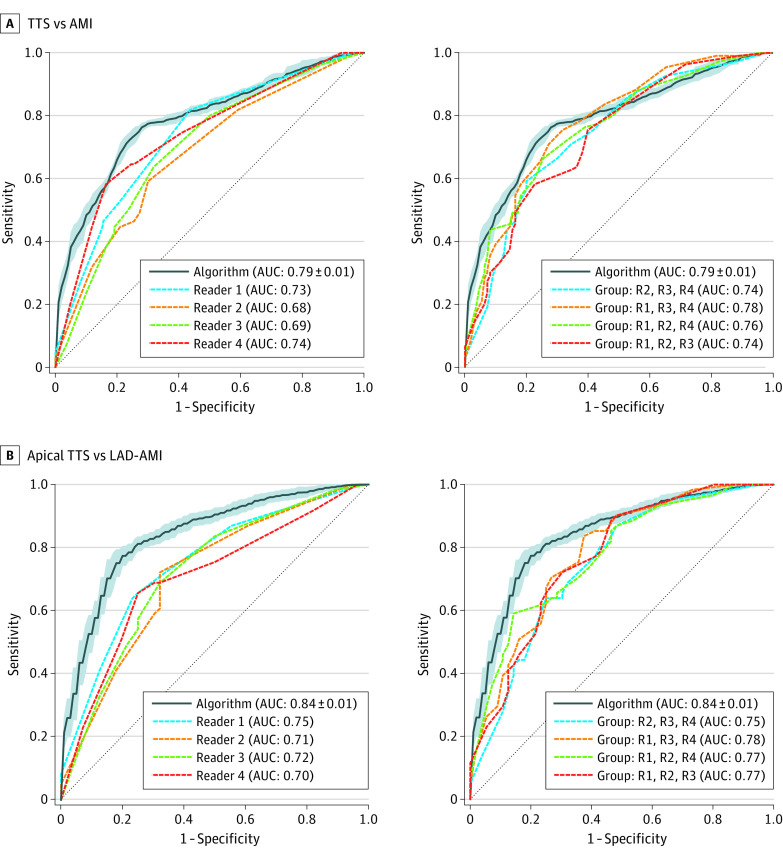
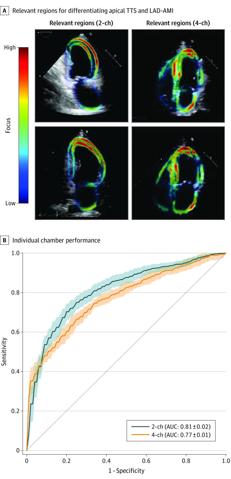
Results: In this cohort study, apical 2-chamber and 4-chamber echocardiographic views of 110 patients with TTS (mean [SD] age, 68.4 [12.1] years; 103 [90.4%] were female) and 110 patients with AMI (mean [SD] age, 69.1 [12.2] years; 103 [90.4%] were female) from an independent data set were evaluated. This approach achieved a mean (SD) AUC of 0.79 (0.01) with an overall accuracy of 74.8 (0.7%). In comparison, cardiologists achieved a mean (SD) AUC of 0.71 (0.03) and accuracy of 64.4 (3.5%) on the same data set. In a subanalysis based on 61 patients with apical TTS and 56 patients with AMI due to occlusion of the left anterior descending coronary artery, the model achieved a mean (SD) AUC score of 0.84 (0.01) and an accuracy of 78.6 (1.6%), outperforming the 4 practicing cardiologists (mean [SD] AUC, 0.72 [0.02]) and accuracy of 66.9 (2.8%).

Conclusions and relevance: In this cohort study, a real-time system for fully automated interpretation of echocardiogram videos was established and trained to differentiate TTS from AMI. While this system was more accurate than cardiologists in echocardiography-based disease classification, further studies are warranted for clinical application.


Conflict of interest statement

Conflict of Interest Disclosures: Dr Laumer reported grants from PHRT - SHFN/SWISSHEART Failure Network with project number 2018-122 during the conduct of the study. Dr Delgado reported grants from Abbott Vascular paid to the department of Cardiology of LUMC, personal fees from Abbott Vascular, grants from Bayer paid to the department of Cardiology of LUMC, grants from Biotronik paid to the department of Cardiology of LUMC, grants from Bioventrix paid to the department of Cardiology of LUMC, grants from Boston Scientific paid to the department of Cardiology of LUMC, grants from Edwards Lifesciences paid to the department of Cardiology of LUMC, personal fees from Edwards Lifesciences, grants from GE Healthcare paid to the department of Cardiology of LUMC, personal fees from GE Healthcare, grants from Ionnis paid to the department of Cardiology of LUMC, grants from Medtronic paid to the department of Cardiology of LUMC, personal fees from Medtronic, personal fees from MSD, and personal fees from Novartis outside the submitted work. Dr Bax reported personal fees from Abbott speaker bureau, personal fees from Edwards Lifesciences speaker bureau, grants from Abbott, grants from Edwards Lifesciences, grants from Medtronic, grants from Boston Scientific, grants from Biotronik, and grants from GE Healthcare outside the submitted work. Dr Ruschitzka reported not receiving personal payments by pharmaceutical companies or device manufacturers in the last 3 years (remuneration for the time spent in activities, such as participation as steering committee member of clinical trials and member of the Pfizer Research Award selection committee in Switzerland, were made directly to the University of Zurich). The Department of Cardiology (University Hospital of Zurich/University of Zurich) reports research-, educational- and/or travel grants from Abbott, Amgen, AstraZeneca, Bayer, Berlin Heart, B. Braun, Biosense Webster, Biosensors Europe AG, Biotronik, BMS, Boehringer Ingelheim, Boston Scientific, Bracco, Cardinal Health Switzerland, Corteria, Daiichi, Diatools AG, Edwards Lifesciences, Guidant Europe NV (BS), Hamilton Health Sciences, Kaneka Corporation, Kantar, Labormedizinisches Zentrum, Medtronic, MSD, Mundipharma Medical Company, Novartis, Novo Nordisk, Orion, Pfizer, Quintiles Switzerland Sarl, Sahajanand IN, Sanofi, Sarstedt AG, Servier, SIS Medical, SSS International Clinical Research, Terumo Deutschland, Trama Solutions, V- Wave, Vascular Medical, Vifor, Wissens Plus, and ZOLL. The research and educational grants do not impact on Dr Ruschitzka’s personal remuneration. Dr Templin reported personal fees from Biotronik Consulting fees, personal fees from Microport Consulting, personal fees from Schnell Medical Consulting, personal fees from Novartis Lecture, and other from Amgen Advisory board outside the submitted work. No other disclosures were reported.

Figures

Figure 1.
Figure 1.. Overview of the Fully Automated Echocardiographic Video Interpretation
The overall method consists of 3 main components: input processing, feature extraction, and sequence classification. First, each frame of echocardiogram video is semantically segmented (A) and the resulting videos are artificially augmented (B). In the next step, an autoencoder model is trained to reconstruct the segmented masks (C). The trained autoencoder is then used to extract a lower dimensional representation of each frame. A multivariate sequence originating from concatenating such representations is depicted along the time axis (D). In the final step, the sequences are artificially augmented and resampled (E), and a temporal neural network based on 1-dimensional time convolutional architecture is trained to classify the sequences according to takotsubo syndrome and acute myocardial infarction (F). CNN indicates convolutional neural network; 2-ch, 2 chamber; 4-ch, 4 chamber.
Figure 2.
Figure 2.. Performance of the Machine Learning Algorithm Compared With Cardiologists
Receiver operating characteristic (ROC) curve (TTS [takotsubo syndrome] considered as the positive class) of the algorithm plotted against the ROC of the 4 different readers. The left of each panel compares the algorithm performance against individual readers. The right of each panel compares the performance with an expert voting committee consisting of 3 of 4 readers. In panel A, the performance of the machine learning algorithm is evaluated on the whole test data set. In panel B, the algorithm is evaluated on a subset of patients with common subtypes, ie, apical TTS and LAD-AMI. AMI indicates acute myocardial infarction, AUC, area under the curve; LAD-AMI, left anterior descending AMI; R, reader.
Figure 3.
Figure 3.. Interpretability Analysis (Apical TTS vs LAD-AMI)
A, The individual latent sequences extracted from a video track the movement of different parts of the myocardium. The colored parts in the ultrasound image correspond to the area on which the latent sequences that performed best in terms of area under the curve (AUC) predominantly focused. The analysis is done separately for the 2-chamber and 4-chamber view. B, The ROC curve based on the 2 different chamber views using all sequences is presented. The SD is calculated based on 5 different training and classification runs of the temporal neural network. LAD-AMI indicates left anterior descending acute myocardial infarction; TTS, takotsubo syndrome; 2-ch, 2 chamber; 4-ch, 4 chamber.

Comment in

References

    1. Krittanawong C, Zhang H, Wang Z, Aydar M, Kitai T. Artificial intelligence in precision cardiovascular medicine. J Am Coll Cardiol. 2017;69(21):2657-2664. doi:10.1016/j.jacc.2017.03.571 - DOI - PubMed
    1. Poplin R, Varadarajan AV, Blumer K, et al. . Prediction of cardiovascular risk factors from retinal fundus photographs via deep learning. Nat Biomed Eng. 2018;2(3):158-164. doi:10.1038/s41551-018-0195-0 - DOI - PubMed
    1. Narula S, Shameer K, Salem Omar AM, Dudley JT, Sengupta PP. Machine-learning algorithms to automate morphological and functional assessments in 2d echocardiography. J Am Coll Cardiol. 2016;68(21):2287-2295. doi:10.1016/j.jacc.2016.08.062 - DOI - PubMed
    1. Sanchez-Martinez S, Duchateau N, Erdei T, et al. . Machine learning analysis of left ventricular function to characterize heart failure with preserved ejection fraction. Circ Cardiovasc Imaging. 2018;11(4):e007138. doi:10.1161/CIRCIMAGING.117.007138 - DOI - PubMed
    1. Zhang J, Gajjala S, Agrawal P, et al. . Fully automated echocardiogram interpretation in clinical practice. Circulation. 2018;138(16):1623-1635. doi:10.1161/CIRCULATIONAHA.118.034338 - DOI - PMC - PubMed

Publication types