. 2022 Jul;52(8):1462-1475.
doi: 10.1007/s00247-022-05327-5. Epub 2022 Mar 30.

Comparison of cardiac volumetry using real-time MRI during free-breathing with standard cine MRI during breath-hold in children

Affiliations

Comparison of cardiac volumetry using real-time MRI during free-breathing with standard cine MRI during breath-hold in children

Lena Maria Röwer et al. Pediatr Radiol. 2022 Jul.

Abstract

Background: Cardiac real-time magnetic resonance imaging (RT-MRI) provides high-quality images even during free-breathing. Difficulties in post-processing impede its use in clinical routine.

Objective: To demonstrate the feasibility of quantitative analysis of cardiac free-breathing RT-MRI and to compare image quality and volumetry during free-breathing RT-MRI in pediatric patients to standard breath-hold cine MRI.

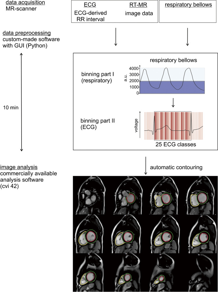
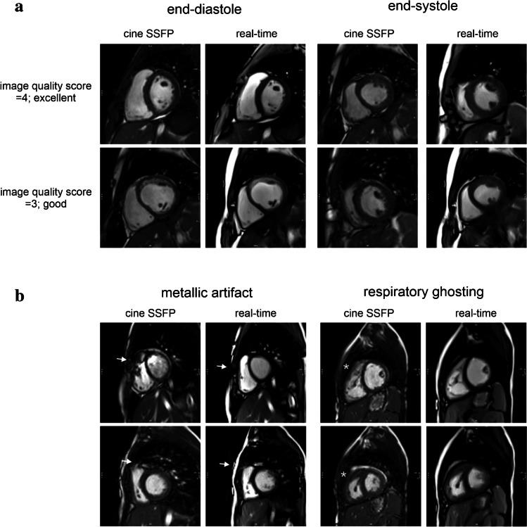
Materials and methods: Pediatric patients (n = 22) received cardiac RT-MRI volumetry during free breathing (1.5 T; short axis; 30 frames per s) in addition to standard breath-hold cine imaging in end-expiration. Real-time images were binned retrospectively based on electrocardiography and respiratory bellows. Image quality and volumetry were compared using the European Cardiovascular Magnetic Resonance registry score, structure visibility rating, linear regression and Bland-Altman analyses.

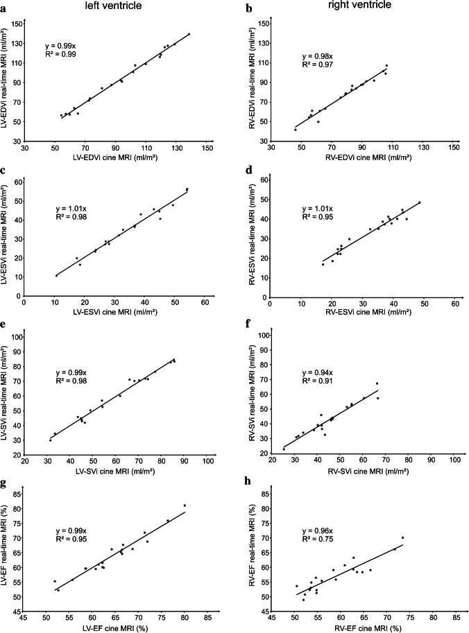
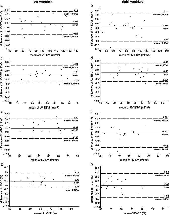
Results: Additional time for binning of real-time images was 2 min. For both techniques, image quality was rated good to excellent. RT-MRI was significantly more robust against artifacts (P < 0.01). Linear regression revealed good correlations for the ventricular volumes. Bland-Altman plots showed a good limit of agreement (LoA) for end-diastolic volume (left ventricle [LV]: LoA -0.1 ± 2.7 ml/m2, right ventricle [RV]: LoA -1.9 ± 3.4 ml/m2), end-systolic volume (LV: LoA 0.4 ± 1.9 ml/m2, RV: LoA 0.6 ± 2.0 ml/m2), stroke volume (LV: LoA -0.5 ± 2.3 ml/m2, RV: LoA -2.6 ± 3.3 ml/m2) and ejection fraction (LV: LoA -0.5 ± 1.6%, RV: LoA -2.1 ± 2.8%).

Conclusion: Compared to standard cine MRI with breath hold, RT-MRI during free breathing with retrospective respiratory binning offers good image quality, reduced image artifacts enabling fast quantitative evaluations of ventricular volumes in clinical practice under physiological conditions.

Keywords: Children; Computer-assisted; Heart; Image processing; Magnetic resonance imaging; Respiration; Volumetry.


Conflict of interest statement

Jens Frahm and Dirk Voit are co-inventors of a patent and software describing the real-time MRI technique used here. The other authors declare no conflicts of interest.

Figures

Fig. 1
Fig. 1
RT-MRI processing. RT images in short axis orientation included the ECG derived time after the R-peak in their DICOM tags. A custom-made software program assigned the respiratory bellows values to the DICOM tags. Images with respiratory bellows values corresponding to a low lung volume (respiratory bellows values < 2000 a.u.) were binned into 25 ECG classes. RT images (from RT balanced SSFP sequence, planes 5-16) were imported into a commercially available analysis software (cvi42) for contouring. a.u. arbitrary units, cvi cardiovascular imaging, DICOM Digital Imaging and Communications in Medicine, ECG electrocardiography, RT real-time, RT-MRI real-time magnetic resonance imaging, SSFP steady-state free precession
Fig. 2
Fig. 2
Image quality analysis. a, b Real time and conventional MR images in short axis orientation. a Standard cine images (conventional cine balanced SSFP sequence, planes 6 and 9) and RT images (RT balanced SSFP sequence, planes 6 and 9) of 7-, 14- and 16-year-old patients no. 4, 12, 18, (female, male, male), were rated good = 3 and excellent = 4 on average for structure visualization based on established 4-point scales [13, 15]. b The most common artifacts were metallic artifacts (e.g., sternal clips) (arrows), which affected standard cine MRI significantly more often and more severely than RT-MRI. Images of 11-year-old patient no. 9 (female). Respiratory artifacts (stars) occurred more often with cine MRI whereas RT-MRI was not affected by respiratory ghosting. Images of 5-year-old patient no. 19 (female). MRI magnetic resonance imaging, RT real-time, RT-MRI real-time magnetic resonance imaging, SSFP steady-state free precession
Fig. 3
Fig. 3
Representative images of the youngest patient, a 5-year-old girl (patient 19) falling asleep during RT-MRI. Images in short axis orientation from basal (plane 8), mid-ventricular (plane 11) and apical (plane 15) levels are presented at end-diastole and end-systole for RT-MRI (RT balanced SSFP sequence) and corresponding cine MRI (conventional cine balanced SSFP sequence). RT-MRI showed excellent image quality. In contrast, cine MRI suffered from respiratory ghosting. Arrows mark respiratory artifacts. MRI magnetic resonance imaging, RT-MRI real-time magnetic resonance imaging, SSFP steady-state free precession
Fig. 4
Fig. 4
Linear regression analysis. Linear regression analyses for LV (a, c, e, g) and RV (b, d, f, h) volumetric measurements in cine MRI and RT-MRI revealed good correlations in EDVi (ml/m2), ESVi (ml/m2), SVi (ml/m2) and EF (%). The regression lines, corresponding linear equations and coefficients of determination are integrated into the graphs. EDVi end-diastolic volume indexed to body surface area, EF ejection fraction, ESVi end-systolic volume indexed to body surface area, LV left ventricle, RT-MRI real-time magnetic resonance imaging, RV right ventricle, MRI magnetic resonance imaging, SVi stroke volume indexed to body surface area
Fig. 5
Fig. 5
Bland–Altman plots. Bland–Altman plots for LV (a, c, e, g) and RV (b, d, f, h) demonstrate the calculated difference (RT-MRI – cine MRI) on the y-axis as a function of the mean value from both imaging techniques on the x-axis. Solid lines represent the mean value of the differences (RT-MRI – cine MRI); dashed lines show the mean difference ± 1.96 standard deviation. LV left ventricle, MRI magnetic resonance imaging, RT-MRI real-time magnetic resonance imaging, RV right ventricle

Similar articles

Cited by

References

    1. Constantine G, Shan K, Flamm SD, Sivananthan MU. Role of MRI in clinical cardiology. Lancet. 2004;363:2162–2171. doi: 10.1016/S0140-6736(04)16509-4. - DOI - PubMed
    1. Deutsch HJ, Smolorz J, Sechtem U, et al. Cardiac function by magnetic resonance imaging. Int J Card Imaging. 1988;3:3–11. doi: 10.1007/BF01801639. - DOI - PubMed
    1. Niwa K, Uchishiba M, Aotsuka H, et al. Measurement of ventricular volumes by cine magnetic resonance imaging in complex congenital heart disease with morphologically abnormal ventricles. Am Heart J. 1996;131:567–575. doi: 10.1016/S0002-8703(96)90538-4. - DOI - PubMed
    1. Matsumura K, Nakase E, Haiyama T, et al. Determination of cardiac ejection fraction and left ventricular volume: contrast-enhanced ultrafast cine MR imaging vs IV digital subtraction ventriculography. AJR Am J Roentgenol. 1993;160:979–985. doi: 10.2214/ajr.160.5.8470613. - DOI - PubMed
    1. Funk E, Thunberg P, Anderzen-Carlsson A. Patients' experiences in magnetic resonance imaging (MRI) and their experiences of breath holding techniques. J Adv Nurs. 2014;70:1880–1890. doi: 10.1111/jan.12351. - DOI - PubMed

LinkOut - more resources