. 2022 Jul;70(7):1301-1316.
doi: 10.1002/glia.24174. Epub 2022 Mar 30.

The two pore potassium channel THIK-1 regulates NLRP3 inflammasome activation

Affiliations

The two pore potassium channel THIK-1 regulates NLRP3 inflammasome activation

Samuel Drinkall et al. Glia. 2022 Jul.

Abstract

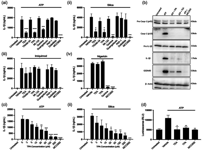
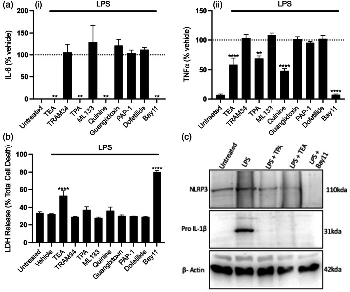
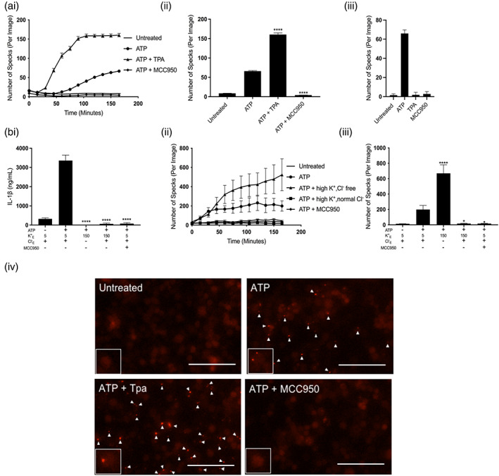
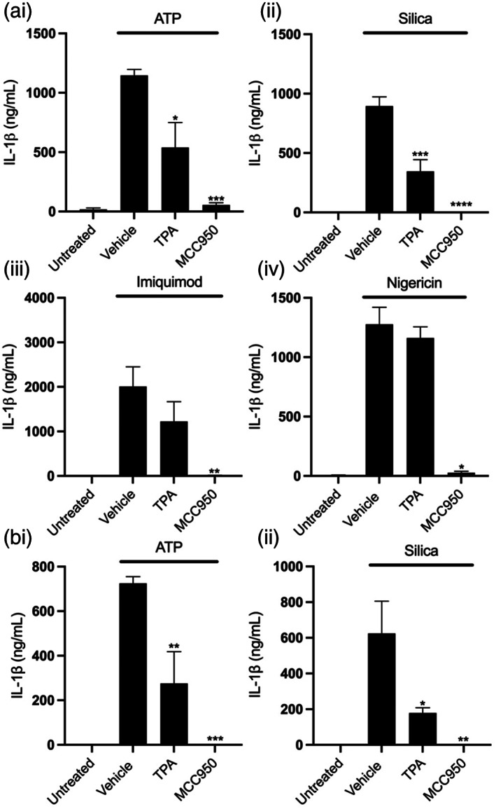
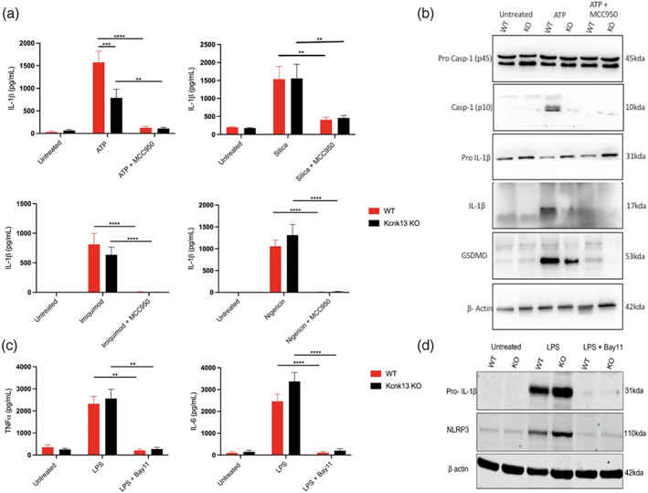
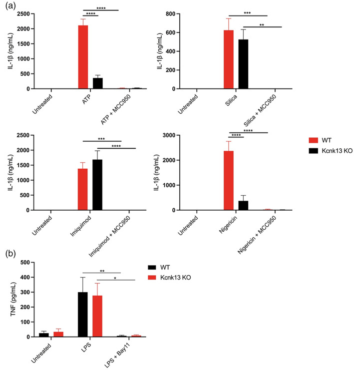
The NLRP3 (NLR family, pyrin domain containing 3) inflammasome is a multi-protein complex responsible for the activation of caspase-1 and the subsequent cleavage and activation of the potent proinflammatory cytokines IL-1β and IL-18, and pyroptotic cell death. NLRP3 is implicated as a driver of inflammation in a range of disorders including neurodegenerative diseases, type 2 diabetes, and atherosclerosis. A commonly reported mechanism contributing to NLRP3 inflammasome activation is potassium ion (K+ ) efflux across the plasma membrane. Identification of K+ channels involved in NLRP3 activation remains incomplete. Here, we investigated the role of the K+ channel THIK-1 in NLRP3 activation. Both pharmacological inhibitors and cells from THIK-1 knockout (KO) mice were used to assess THIK-1 contribution to macrophage NLRP3 activation in vitro. Pharmacological inhibition of THIK-1 inhibited caspase-1 activation and IL-1β release from mouse bone-marrow-derived macrophages (BMDMs), mixed glia, and microglia in response to NLRP3 agonists. Similarly, BMDMs and microglia from THIK-1 KO mice had reduced NLRP3-dependent IL-1β release in response to P2X7 receptor activation with ATP. Overall, these data suggest that THIK-1 is a regulator of NLRP3 inflammasome activation in response to ATP and identify THIK-1 as a potential therapeutic target for inflammatory disease.

Keywords: NLRP3; THIK-1; inflammasome; inflammation; interleukin-1; potassium channel.


Figures

FIGURE 1
FIGURE 1
Pharmacological inhibition of two pore domain potassium channels blocks NLRP3 inflammasome activation and IL‐1β processing. (ai–iii) IL‐1β ELISA of the supernatant of pBMDMs primed with LPS (1 μg ml−1, 4 h) followed by pretreatment with MCC950 (10 μM) or K+ channel inhibitors TEA (50 mM), TRAM‐34 (10 μM), TPA (50 μM), ML‐133 (20 μM), quinine (100 μM), Guangitoxin‐1E (25 nM), PAP‐1 (2 μM), or Dofetilide (1 μM) for 15 min before stimulation with ATP (5 mM, 1 h) (n = 4), silica (300 μg ml−1, 4 h) (n = 5) or imiquimod (75 μM, 2 h) (n = 3). (Aiv) IL‐1β ELISA of the supernatant of pBMDMs primed with LPS (1 μg ml−1, 4 h) followed by pretreatment with MCC950 (10 μM) or TEA (50 mM), TPA (50 μM), quinine (100 μM), for 15 min before stimulation with nigericin (10 μM, 1 h) (n = 4). (b) Caspase‐1, IL‐ β and gasdermin D western blot of total cell lysates (cell lysate + supernatant) from LPS‐primed (1 μg ml−1, 4 h) pBMDMs pretreated with vehicle control, TEA (50 mM), TPA (50 μM) or MCC950 (10 μM) for 15 min then stimulated with ATP (5 mM, 1 h). (c) IL‐1β ELISA of the supernatant of pBMDMs primed with LPS (1 μg ml−1, 4 h) followed by pretreatment with MCC950 (10 μM) or TPA (3–300 μM) before stimulation with ATP (5 mM, 1 h) (n = 5) or silica (300 μg ml−1, 4 h) (n = 3). (d) Caspase‐1 Glo assay to measure caspase‐1 activity of LPS‐primed (1 μg ml−1, 4 h) pBMDMs pretreated with vehicle control, TEA (50 mM), TPA (50 μM) or MCC950 (10 μM) for 15 min then stimulated with ATP (5 mM, 1 h). ****p < .0001, ***p < .001, **p < .01, *p < .05 determined by one‐way ANOVA with Dunnett's post hoc analysis. Values shown are the mean ± SEM
FIGURE 2
FIGURE 2
Pharmacological inhibition of two pore potassium channels blocks priming of the NLRP3 inflammasome. (a) IL‐6 (i) and TNF (ii) ELISA and (b) LDH release assay of the supernatant of iBMDMs pretreated with Bay11(10 μM) or K+ channel inhibitors TEA (50 mM), TRAM‐34 (10 μM), TPA (50 μM), ML‐133 (20 μM), quinine (100 μM), Guangitoxin‐1E (25 nM), PAP‐1 (2 μM) or Dofetilide (1 μM) for 15 min before priming with LPS (1 μg ml−1, 4 h) (n = 4). (c) NLRP3 and IL‐1β western blot of the supernatant and total cell lysates respectively of iBMDMs pretreated with TEA (50 mM), TPA (50 μM) or Bay11 (10 μM) for 15 min before priming with LPS (1 μg ml−1, 4 h). ****p < .0001, **p < .01 determined by one‐way ANOVA with Dunnett's post hoc analysis. Values shown are the mean ± SEM
FIGURE 3
FIGURE 3
Potassium efflux is required for NLRP3 inflammasome activation but not ASC speck formation in response to ATP. (a, i) ASC speck formation measured in real time and (a, ii) ASC speck formation after 165 min of ATP stimulation from ASC‐mCherry iBMDMs primed with LPS (1 μg ml−1, 4 h) followed by pretreatment vehicle control, TPA (50 μM) or MCC950 (10 μM) for 15 min before stimulation with ATP (5 mM) (n = 6). (a, iii) ASC speck formation after 165 min from ASC‐mCherry iBMDMS primed with LPS (1 μg ml−1, 4 h) followed by treatment with vehicle control, TPA (50 μM) or MCC950 (10 μM) in the absence of ATP (n = 6). (b, i) IL‐1β ELISA of the supernatant of iBMDMs primed with LPS (1 μg ml−1, 4 h) followed by incubation in a control (145 mM NaCl/ 5 mM KCl), high K+ and normal Cl (150 mM KCl), high K+ and Cl free (150 mM KGluconate) or control and MCC950 (10 μM) solution for 15 min before stimulation with ATP (5 mM, 1 h) (n = 6). (b, ii) ASC speck formation measured in real time and (b, iii) ASC speck formation after 165 min of ATP stimulation from iBMDMs stably expressing ASC‐mCherry (ASC‐mCherry iBMDMs) primed with LPS (1 μg ml−1, 4 h) followed by incubation in a control (145 mM NaCl/ 5 mM KCl), high K+ and normal Cl (150 mM KCl), high K+ and Cl free (150 mM KGluconate) or control and MCC950 (10 μM) solution for 15 min before stimulation with ATP (5 mM) (n = 4). (b, iv) representative images of ASC‐mCherry iBMDMs after 165 min ATP stimulation (scale bar, 50 μm, arrows denote ASC specks). ASC speck experiments were performed in the presence of ac‐YVAD‐CMK (50 μM) to prevent pyroptosis and loss of ASC specks. ****p < .0001, *p < .05 determined by one‐way ANOVA with Dunnett's post hoc analysis. Values shown are the mean ± SEM
FIGURE 4
FIGURE 4
Inhibition of THIK‐1 blocks NLRP3 activation in mixed glia and isolated microglia. (a) IL‐1β ELISA of the supernatant of primary mouse mixed glia primed with LPS (1 μg ml−1, 4 h) followed by pretreatment with vehicle control, TPA (50 μM) or MCC950 (10 μM) for 15 min before stimulation with ATP (5 mM, 1 h) (n = 5), silica (300 μg ml−1,4 h) (n = 3), imiquimod (75 μM, 2 h) (n = 4) or nigericin (10 μM, 1 h) (n = 3). (b) IL‐1β ELISA of the supernatant of mouse primary microglia primed with LPS (1 μg ml−1, 4 h) followed by pretreatment with vehicle control, TPA (50 μM) or MCC950 (10 μM) for 15 min before stimulation with ATP (5 mM, 1 h) or silica (300 μg ml−1,4 h) (n = 3). ****p < .0001, ***p < .001, **p < .01, *p < .05 determined by one‐way ANOVA with Dunnett's post hoc analysis. Values shown are the mean ± SEM
FIGURE 5
FIGURE 5
THIK‐1 specifically regulates ATP‐induced NLRP3 activation in bone‐marrow‐derived macrophages. (a) IL‐1β ELISA of the supernatant of primary wild‐type (WT) and Kcnk13 knockout (KO) BMDMs primed with LPS (1 μg ml−1, 4 h) followed by pretreatment with MCC950 (10 μM) for 15 min before stimulation with ATP (5 mM, 1 h) (n = 7), silica (300 μg ml−1,4 h) (n = 7), imiquimod (75 μM, 2 h) (n = 4) or nigericin (10 μM, 1 h) (n = 8). (b) Caspase‐1, IL‐1β and gasdermin D western blot of total cell lysates (cell lysate + supernatant) from LPS‐primed WT and Kcnk13 KO pBMDMs pretreated with vehicle control or MCC950 (10 μM) for 15 min before stimulated with ATP (5 mM, 1 h). (c and d) TNF and IL‐6 ELISA and NLRP3 and IL‐1β western blot of the supernatant and total cell lysates respectively of primary wild‐type (WT) and Kcnk13 knockout (KO) BMDMs pretreated with Bay11(10 μM) for 15 min before priming with LPS (1 μg ml−1, 4 h) (n = 7). ****p < .0001, ***p < .001, **p < .01 determined by two‐way ANOVA with Bonferroni's post hoc analysis. Values shown are the mean ± SEM
FIGURE 6
FIGURE 6
THIK‐1 regulates NLRP3 activation in primary adult microglia. (a) IL‐1β ELISA of the supernatant of primary wild‐type (WT) and Kcnk13 knockout (KO) adult microglia primed with LPS (1 μg ml−1, 4 h) followed by pretreatment with MCC950 (10 μM) for 15 min before stimulation with ATP (5 mM, 1 h) (n = 4), silica (300 μg ml−1,4 h) (n = 3), imiquimod (75 μM, 2 h) (n = 3) or nigericin (10 μM, 1 h) (n = 4). (b) TNF ELISA of the supernatant of primary wild‐type (WT) and Kcnk13 knockout (KO) primary adult microglia pretreated with Bay11(10 μM) for 15 min before priming with LPS (1 μg ml−1, 4 h) (n = 5). ****p < .0001, ***p < .001, **p < .01, *p < .05 determined by two‐way ANOVA with Bonferroni's post hoc analysis. Values shown are the mean ± SEM

References

    1. Akopova, O. V. (2017). Direct and off‐target effects of ATP‐sensitive potassium channels opener diazoxide. Journal of Drug Metabolism & Toxicology, 8, 227. 10.4172/2157-7609.1000227 - DOI
    1. Baldwin, A. G. , Rivers‐Auty, J. , Daniels, M. J. D. , White, C. S. , Schwalbe, C. H. , Schilling, T. , Hammadi, H. , Jaiyong, P. , Spencer, N. G. , England, H. , Luheshi, N. M. , Kadirvel, M. , Lawrence, C. B. , Rothwell, N. J. , Harte, M. K. , Bryce, R. A. , Allan, S. M. , Eder, C. , Freeman, S. , & Brough, D. (2017). Boron‐based inhibitors of the NLRP3 Inflammasome. Cell Chemical Biology, 24, 1321–1335. 10.1016/j.chembiol.2017.08.011 - DOI - PMC - PubMed
    1. Baron, L. , Gombault, A. , Fanny, M. , Villeret, B. , Savigny, F. , Guillou, N. , Panek, C. , Le Bert, M. , Lagente, V. , Rassendren, F. , Riteau, N. , & Couillin, I. (2015). The NLRP3 inflammasome is activated by nanoparticles through ATP, ADP and adenosine. Cell Death and Disease, 6, e1629. 10.1038/cddis.2014.576 - DOI - PMC - PubMed
    1. Bauernfeind, F. , Horvath, G. , Stutz, A. , Alnemri, E. S. , Speert, D. , Fernandes‐alnemri, T. , Wu, J. , Brian, G. , Fitzgerald, K. A. , Hornung, V. , & Latz, E. (2009). NF‐kB activating pattern recognition and cytokine receptors license NLRP3 inflammasome activation by regulating NLRP3 expression. Journal of Immunology, 183, 787–791. - PMC - PubMed
    1. Bittner, S. , Bobak, N. , Herrmann, A. M. , Göbel, K. , Meuth, P. , Höhn, K. G. , Stenner, M. P. , Budde, T. , Wiendl, H. , & Meuth, S. G. (2010). Upregulation of K2P5.1 potassium channels in multiple sclerosis. Annals of Neurology, 68, 58–69. 10.1002/ana.22010 - DOI - PubMed

Publication types

MeSH terms

LinkOut - more resources