. 2022 Apr 13;144(14):6326-6342.
doi: 10.1021/jacs.1c13568. Epub 2022 Mar 30.

Covalent Proximity Scanning of a Distal Cysteine to Target PI3Kα

Affiliations

Covalent Proximity Scanning of a Distal Cysteine to Target PI3Kα

Chiara Borsari et al. J Am Chem Soc. .

Abstract

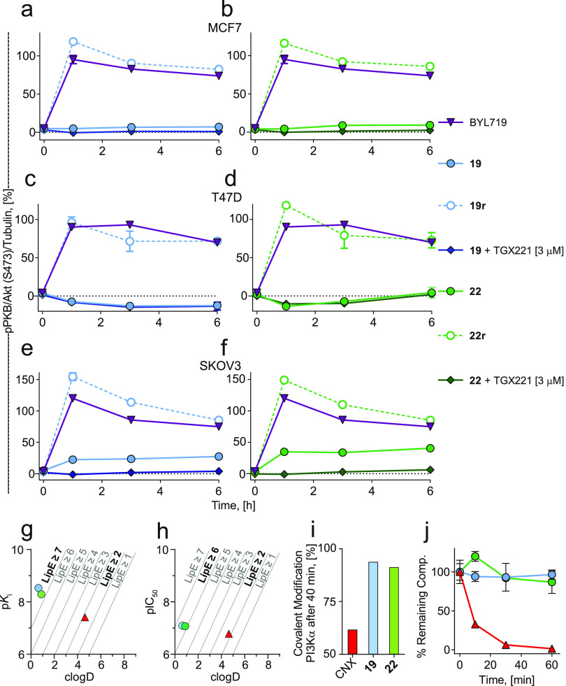
Covalent protein kinase inhibitors exploit currently noncatalytic cysteines in the adenosine 5'-triphosphate (ATP)-binding site via electrophiles directly appended to a reversible-inhibitor scaffold. Here, we delineate a path to target solvent-exposed cysteines at a distance >10 Å from an ATP-site-directed core module and produce potent covalent phosphoinositide 3-kinase α (PI3Kα) inhibitors. First, reactive warheads are used to reach out to Cys862 on PI3Kα, and second, enones are replaced with druglike warheads while linkers are optimized. The systematic investigation of intrinsic warhead reactivity (kchem), rate of covalent bond formation and proximity (kinact and reaction space volume Vr), and integration of structure data, kinetic and structural modeling, led to the guided identification of high-quality, covalent chemical probes. A novel stochastic approach provided direct access to the calculation of overall reaction rates as a function of kchem, kinact, Ki, and Vr, which was validated with compounds with varied linker lengths. X-ray crystallography, protein mass spectrometry (MS), and NanoBRET assays confirmed covalent bond formation of the acrylamide warhead and Cys862. In rat liver microsomes, compounds 19 and 22 outperformed the rapidly metabolized CNX-1351, the only known PI3Kα irreversible inhibitor. Washout experiments in cancer cell lines with mutated, constitutively activated PI3Kα showed a long-lasting inhibition of PI3Kα. In SKOV3 cells, compounds 19 and 22 revealed PI3Kβ-dependent signaling, which was sensitive to TGX221. Compounds 19 and 22 thus qualify as specific chemical probes to explore PI3Kα-selective signaling branches. The proposed approach is generally suited to develop covalent tools targeting distal, unexplored Cys residues in biologically active enzymes.


Conflict of interest statement

The authors declare no competing financial interest.

Figures

Figure 1
Figure 1
(a, c) Design strategy for the development of irreversible inhibitors targeting (a) BTK and (c) EGFR. X-ray crystallographic structure of (a) BTK (colored wheat) in complex with PCI-29732 (teal; PDB ID 3GEN) and (c) EGFR (cyan) in complex with gefitinib (brown, PDB ID 4I22). (b, d) Chemical structure of the reversible-inhibitor scaffolds and covalent derivatives are depicted. The incorporation of the warhead on PCI-29732 led to acabrutinib and ibrutinib, and gefitinib served as a scaffold for dacomitinib and afatinib. (e) PQR514 (magenta) bound to phosphoinositide 3-kinase α (PI3Kα) (gray; based on PDB ID 6OAC(59)). Cys862 positions from eight superimposed PI3Kα-inhibitor complexes are shown (see Table S1 for details). The position of the cysteine thiols (yellow) is conserved across the different complexes and deviates less than 2.6 Å. (f) Schematic design of the covalent PI3Kαi library. The distances between reversible inhibitor skeletons and the targeted nucleophiles are shown as dashed green lines (∼4 Å for BTK and EGFR vs ∼11 Å for PI3Kα).
Figure 2
Figure 2
(a) Chemical structure of a set of nine warhead-containing compounds (19). (b) General reaction of warhead-containing compounds with βME. (c) High-performance liquid chromatography (HPLC) reaction monitoring of inhibitor (3) and its βME adduct (3-βME) formation. (d) Time-dependent inhibitor consumption curves used to calculate kchem. Values are mean ± standard deviation (SD) (n = 3). Error bars are not shown when smaller than symbols. (e) Chemical shift of the α-proton in 1H NMR spectra of compounds bearing an unsubstituted double bond (7) and β-methyl (8)- and β,β-dimethyl (9)-substituted warheads. (f) Intrinsic reactivity of the inhibitors (kchem) plotted against the chemical shift of the α-protons of the indicated compounds. Raw data and details for kchem calculations are reported in Table S2 and in the Materials and Methods section in the Supporting Information. The second-order rate constant kchem could not be measured for compounds 6 and 9 due to lack of reactivity with 12 M βME, and also not for 1 due to its extremely fast reaction with 1 mM βME. kchem for compound 8 was lower than 1 × 10⁻⁶ M–1·s–1 and was thus considered nonreactive. These three values are therefore “out-off scale” (denoted with red arrows) for kchem. (g) Representation of energy of the lowest-unoccupied molecular orbital (LUMO) electron density (top) and LUMO map (bottom) for compound 1. Electron deficiency ranges from blue (high) to red (low). Given that the LUMO designates space available for a pair of electrons, color tints toward blue in the LUMO map indicate where a nucleophilic attack would likely occur. Calculations were performed using Spartan 18, Wavefunction, Inc. (h) ELUMO values plotted against the intrinsic reactivity of the inhibitors (kchem). Raw data and details for kchem calculations are reported in Table S2 and in the Materials and Methods section in the Supporting Information. As in (f), values for kchem denoted with red arrows are “out-off scale”. (i) Chemical shift of α-protons of indicated compounds plotted against ELUMO values. (j) Model for cellular on- and off-target reactions of covalent inhibitors. In the on-target reaction (green), enzyme E and inhibitor I form first a reversible E∼I complex (equilibrium dependent on Ki), which is then converted to a covalent EI complex (controlled by kinact). Competing off-target reactions (red) with cellular sulfhydryls (S) consume the inhibitor and form a stable SI adduct. (k) On- and off-target covalent reactions of inhibitors as depicted in (j) were modeled using KinTek Global Kinetic Explorer: concentrations were set to 7 mM for intracellular sulfhydryls (for intracellular reduced glutathione (GSH) concentrations, see ref (80)); for the targeted enzyme (PI3Kα) to 10 nM; and for indicated covalent inhibitors to 100 nM. Experimental values for kinact and kchem used for the modeling are listed in Table S2 and Figure S1. Details for the determination of kinact are given in Figure 3 and associated materials.
Figure 3
Figure 3
(a) Chemical structure of a collection of nine compounds with pairwise matched linkers and either fast (2, 1012) or moderately (3, 1315a) reactive Michael acceptors, and of 15b and 15c. (b) The reactive volume (Vr) of the warhead was estimated by a sphere around the sulfur atom of Cys862, with a radius equal to the distance (d) between the Cys862 sulfur and the β-carbon of the Michael acceptor (Vr = 4/3·π·d3); see Table S4 for values. The modeled locations of the β-carbon of the Michael acceptor are indicated by spheres colored as in (a). (c) Correlation between kinact and the local warhead concentration (see Table S4 for calculation). (d, e) Time-dependent IC50 determinations derived from time- and concentration-dependent time-resolved fluorescence resonance energy transfer (TR-FRET) ratios curves depicted in Figure S2 (IC50s for all compounds with error bars, see Figure S13). Strong (2, 1012 (d)) and weak (3, 1315a (e)) electrophiles were investigated. (f) Compound-specific intrinsic reactivity (kchem) determined as in Figure 2; (g) dissociation constants (Ki) calculated from concentration and time-dependent TR-FRET experiments as shown in Figure S2 (h) rate constants for covalent binding to PI3Kα (kinact); (i) second-order rate constant used to characterize covalent binding of irreversible inhibitors to the target protein (kinact/Ki). The calculation of kinetic parameters was carried out through global fitting for numerical integration to a kinetic model using KinTek Global Kinetic Explorer modeling software.−,, All values are shown as mean ± SD (n = 3). Error bars are not shown when smaller than the symbols. Orange, strong electrophiles; green, weak electrophiles. The distance between the Michael acceptor (β-carbon) and Cys862 thiol was calculated using PyMOL 2.3.5 Schrödinger, Inc. (see Figure S3 and Table S3). (j) TR-FRET experiments for the reversible analogue of compound 2 (2r). (k) Covalent docking of compounds 2, 1012 using CovDock Schrödinger. Representation of the shift of the morpholine oxygen (spheres, in hinge region defined by Val851) and of the NH2 (spheres, in binding affinity region defined by Asp810) with respect to PQR530 scaffold (magenta) is displayed. (l) CovDock Schrödinger modeling has been used to investigate the displacement of the PI3K core module of the indicated compounds when covalently bound to Cys862 of PI3Kα. The distances of (i) the morpholine oxygen atom (O17) and the N nitrogen atom of Val851 backbone amide (O17-V851, red) in the hinge region, and (ii) the amino group (N26) of the aminopyrimidine and the closest oxygen atom in COO side chain of Asp810 (N26-D810, light blue) in the affinity region were determined from CovDock modeling coordinates from 50 predicted poses, and average and SD were calculated. As a control for a structural distortion of the PI3K-binding module, the distances between the O17 and N26 atoms (O17-N26, black) were determined. Numbering of inhibitor atoms can be found in (a) and (k). Distances are reported in angstrom. Modeling was performed using PDB ID 6OAC(59) as a starting point. All compounds are depicted in Figure S6.
Figure 4
Figure 4
(a) Library of acrylamide-containing compounds (1624). (b) Intrinsic reactivity (kchem) of compounds 1624, ibrutinib and CNX-1351. Experiments performed with HPLC (1 mM inhibitor and 600 mM βME, n = 3). All values are reported as mean ± SD. Error bars are not shown when smaller than the symbols. (c) Efficiency in covalent bond formation (kinact/Ki) plotted against distance from Cys862. Zero on the x-axis corresponds to Cys862 positioning; zero to left: shorter linkers; zero to right: longer linkers. Kinetic parameters are reported in Table S5. The distances between the Michael acceptors (β-carbons) and Cys862 thiol were calculated using Maestro 11.1 and PyMOL 2.3.5 Schrödinger LLC (see Figure S5). (d) Plot of kchem/kinact ratios vs the reactive volume Vr of compounds 12, 2, 11, 10 (red circles), 15a, 3, 14, 13 (green squares), 19, 22, 18, 17, 16 (blue circles), and 20, 23, 21, 24 (orange diamonds). The top x-axis shows the Vr·NA term utilized as in eq S14b to determine the scaling factor σ for each compound group as shown in Figure S7 and Table S6. The compound group-specific σ factors were used to normalize the kchem/kinact ratios by 1/σ. The (blue) regression line includes all compounds listed above except for 20, 23, 21, 24, with a linker reaching beyond the target Cys862. (e) Normalized kcat reaction rates were calculated according to eq 15 with a spherical Vr approximation using the modeled distance to the target d. For Ki, the indicated values were used. (f) Normalized kcat reaction rates were calculated according to eq 17a as a function of d and the inhibitor concentration [I]. For both graphs in (e) and (f), a mean kchem averaged over the values from compounds 19, 22, 18, 17, and 16 was used (kchem (1922181716) = 3.38 × 10⁻⁴ M–1·s–1). (g) LC-SRM quantification of Cys862-modification by 19, 22, and 22r. (h, i) X-ray structure of (h) 19 (deep teal) bound to PI3Kα (PDB ID 7R9V, resolution 2.69 Å) and (i) 22 (deep purple) bound to PI3Kα (PDB ID 7R9Y, resolution 2.85 Å). H-bonds are depicted as dashed black lines. As for the CHF2-group, a H-bond either with (i) Lys802 or with (ii) the triazine core (intramolecular interaction) is possible based on the proton positioning. (j, k) Electron density maps of the inhibitor (2Fo–Fc map contoured at 1σ, blue mesh) fit the structure of (j) 19 and (k) 22, and the electron density clearly shows that the acrylamide forms a covalent bond with the thiol group of Cys862.
Figure 5
Figure 5
(a–d) Residence time experiment using BRET in intact HEK293 cells expressing NanoLuc fused to (a) PI3Kα, (b) PI3Kα Cys862Ser, (c) PI3Kβ, and (d) PI3Kδ. The cells were incubated with inhibitors (3 μM) for 2 h. Subsequently, free inhibitor was washed out twice using Opti-MEM medium (for 10 min at 37 °C and 5% CO2), before the cell-permeable BRET tracer was added (0.2 μM final concentration). A tracer bearing a pyrrolylBODIPY fluorescent moiety (excitation at 460 nm and emission at 618 nm) was used to determine the on-target residence time of the inhibitors after drug washout. Displacement of the inhibitors by the BRET tracer from the ATP-binding pocket in the indicated PI3K isoforms was monitored by the recovery of the BRET signal between the NanoLuc-fused PI3K (donor) and the BRET tracer (acceptor). A prolonged residence time of inhibitors (after drug washout) diminishes the BRET signal and points to a covalent interaction. Data shown are mean ± standard error of the mean (SEM) (n = 3). Results for CNX-1351 are shown in Figure S10.
Figure 6
Figure 6
(a–f) Cellular washout experiments in cancer cells with a mutated and constitutively activated PI3Kα. (a, b) MCF7 (PIK3CA E545K), (c, d) T47D (PIK3CA H1047R), and (e, f) SKOV3 (PIK3CA H1047R; HER2+) cancer cell lines were treated with 3 μM of the indicated compounds for 2 h, followed by drug washout (twice with fully supplemented Dulbecco’s modified Eagle’s medium (DMEM) for 10 min each). The cells were then incubated at 37 °C and 5% CO2 for the indicated times before they were fixed, and the phosphorylation status of PKB/Akt was determined by in-cell western; t = 0 values indicate reference measurements in the presence of inhibitor (i.e., immediately after the 2 h treatment window and without removal of drug). Where indicated, TGX221 (3 μM) was added 1 h before the fixation of cells, to investigate a potential cell-line-dependent further contribution of PI3Kβ to PKB/Akt phosphorylation. Data shown are mean ± SEM (n = 4; two independent measurements). Error bars are not shown when smaller than symbols. (g, h) Plot of clog D vs biological activity ((g) pKi; (h) pIC50) of compounds 19, 22, and CNX-1351. Lipophilic ligand efficiency (LipE) values higher than 5 are considered to be the threshold for lead compounds and high-quality chemical probes. LipE calculations are shown in Table S9. (i) On-target covalent modification modeled after 40 min using KinTek Global Kinetic Explorer. See Figure S1c,d for additional details. (j) Metabolic stability of 19, 22, and CNX-1351 using rat liver microsomes fortified with phase I metabolism cofactor NADPH. The time-dependent degradation of test items (starting concentration 1 μM) in the presence of rat liver microsomes (0.5 mg microsomal protein/mL) was measured (mean ± SD; n = 2). Error bars are not shown when smaller than symbols. Corresponding raw data are reported in Table S10.
Figure 7
Figure 7
(a) Analysis of putative off-target reactions of covalent PI3Kα inhibitors with protein kinases. Coordinates for a cysteinome protein kinase template were kindly provided by Chaikuad and Knapp. Their template was generated based on the PDB ID 1ATP of the catalytic subunit of cAMP-dependent protein kinase (PKA), onto which cysteines throughout the kinome have been projected as reported in ref (70). These coordinates were aligned with PI3K crystallographic data from PDB IDs 5OQ4 (PQR309/PI3Kγ complex), 1E8X (ATP/PI3Kγ complex), and structures of compound 19 bound to PI3Kα provided here, to dock 19 into the PKA template’s ATP-binding site. In the depicted compound 19 (dark blue), the sulfur atom of Cys862 (oversize yellow sphere) is located at a distance of 8.0 Å from Cys F3 (Cys annotation as in ref (70)), which corresponds to a Cys in JNK1, 2, and 3. Closer is only Cys F4 at a distance of 4.0 Å, which is the Cys862 of PI3Kα projected onto the PKA template. The rest of the projected protein kinase Cys residues are ≫10 Å away from the warhead of 19 and cannot be targeted without displacement of the ATP-binding module. (b) Validation of off-target binding of 19 at 1 μM to protein kinases in KINOMEScan experiments (comparisons with results from PQR514, PQR309, BYL719, GDC-0980, and PKI-587, and lipid kinase interactions are shown in Figure S11 and quantitative data are listed in Table S13). The three red spots in the kinase tree represent CSF1R (autoinhibited; juxtamembrane domains of some protein tyrosine kinase receptors stabilize the kinase domain in an inactive state), JAK1 (JH2 domain-pseudokinase), and KIT (autoinhibited) with remaining binding of 20, 32, and 35%, respectively. CSF1R, JAK1, and KIT (and mutant) catalytic domains (in active conformations) showed no relevant interactions with 19.

Similar articles

Cited by

References

    1. Miller V. A.; Hirsh V.; Cadranel J.; Chen Y.-M.; Park K.; Kim S.-W.; Zhou C.; Su W.-C.; Wang M.; Sun Y.; Heo D. S.; Crino L.; Tan E.-H.; Chao T.-Y.; Shahidi M.; Cong X. J.; Lorence R. M.; Yang J. C.-H. Afatinib versus placebo for patients with advanced, metastatic non-small-cell lung cancer after failure of erlotinib, gefitinib, or both, and one or two lines of chemotherapy (LUX-Lung 1): a phase 2b/3 randomised trial. Lancet Oncol. 2012, 13, 528–538. 10.1016/S1470-2045(12)70087-6. - DOI - PubMed
    1. Pan Z.; Scheerens H.; Li S.-J.; Schultz B. E.; Sprengeler P. A.; Burrill L. C.; Mendonca R. V.; Sweeney M. D.; Scott K. C. K.; Grothaus P. G.; Jeffery D. A.; Spoerke J. M.; Honigberg L. A.; Young P. R.; Dalrymple S. A.; Palmer J. T. Discovery of Selective Irreversible Inhibitors for Bruton’s Tyrosine Kinase. ChemMedChem 2007, 2, 58–61. 10.1002/cmdc.200600221. - DOI - PubMed
    1. Yver A. Osimertinib (AZD9291)—a science-driven, collaborative approach to rapid drug design and development. Ann. Oncol. 2016, 27, 1165–1170. 10.1093/annonc/mdw129. - DOI - PubMed
    1. Miles J.; White Y. Neratinib for the Treatment of Early-Stage HER2-Positive Breast Cancer. J. Adv. Pract. Oncol. 2018, 9, 750–754. - PMC - PubMed
    1. Lau S. C. M.; Batra U.; Mok T. S. K.; Loong H. H. Dacomitinib in the Management of Advanced Non-Small-Cell Lung Cancer. Drugs 2019, 79, 823–831. 10.1007/s40265-019-01115-y. - DOI - PubMed

Publication types