. 2022 Apr;8(13):eabj8658.
doi: 10.1126/sciadv.abj8658. Epub 2022 Mar 30.

Neuronal C/EBPβ/AEP pathway shortens life span via selective GABAnergic neuronal degeneration by FOXO repression

Affiliations

Neuronal C/EBPβ/AEP pathway shortens life span via selective GABAnergic neuronal degeneration by FOXO repression

Yiyuan Xia et al. Sci Adv. 2022 Apr.

Abstract

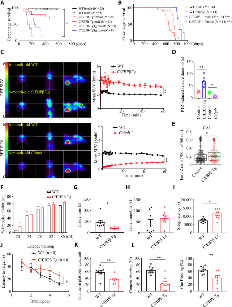
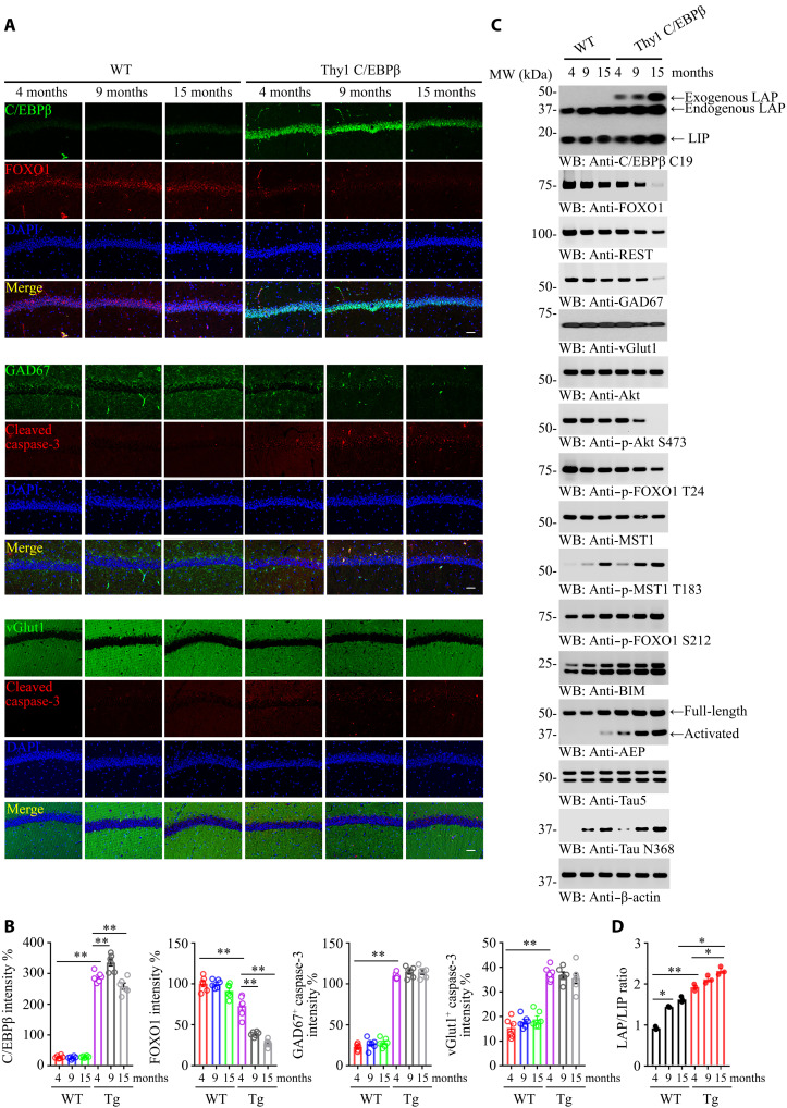
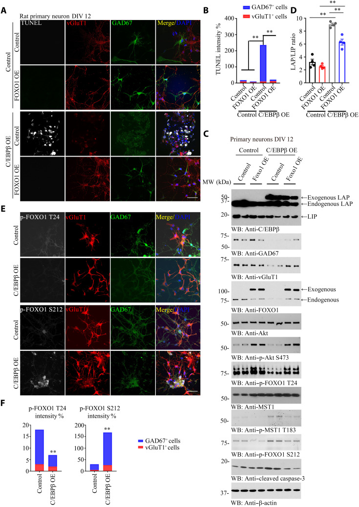
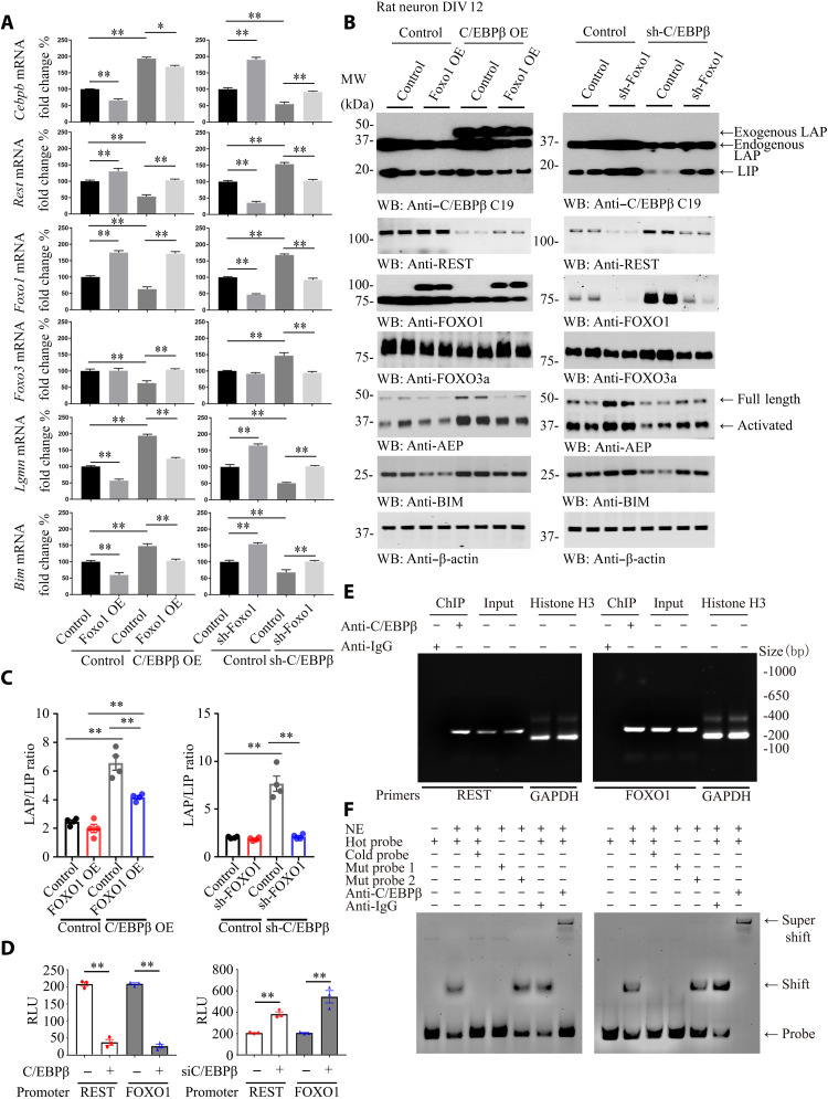
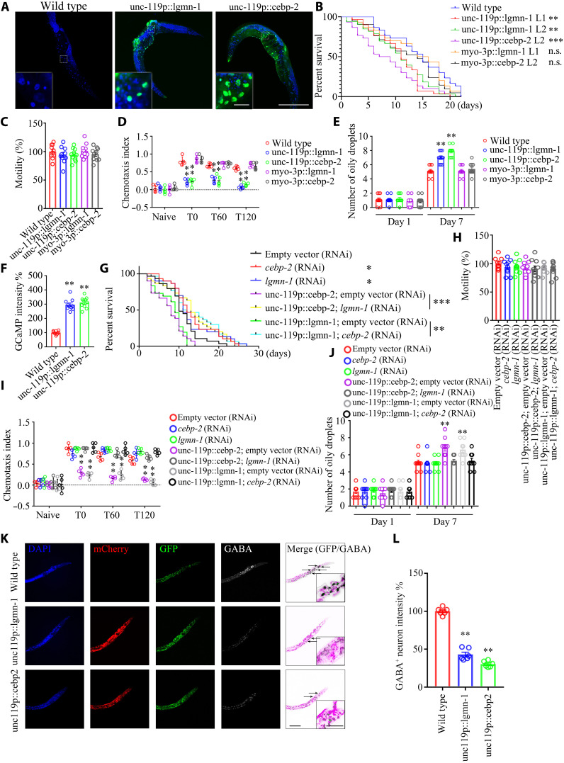
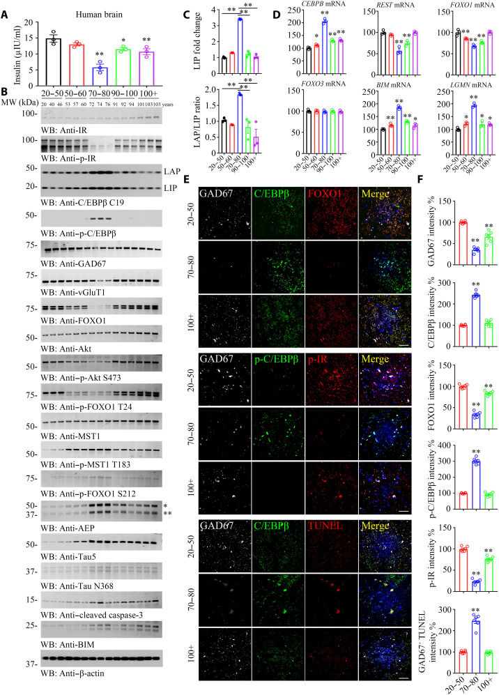
The age-related cognitive decline of normal aging is exacerbated in neurodegenerative diseases including Alzheimer's disease (AD). However, it remains unclear whether age-related cognitive regulators in AD pathologies contribute to life span. Here, we show that C/EBPβ, an Aβ and inflammatory cytokine-activated transcription factor that promotes AD pathologies via activating asparagine endopeptidase (AEP), mediates longevity in a gene dose-dependent manner in neuronal C/EBPβ transgenic mice. C/EBPβ selectively triggers inhibitory GABAnergic neuronal degeneration by repressing FOXOs and up-regulating AEP, leading to aberrant neural excitation and cognitive dysfunction. Overexpression of CEBP-2 or LGMN-1 (AEP) in Caenorhabditis elegans neurons but not muscle stimulates neural excitation and shortens life span. CEBP-2 or LGMN-1 reduces daf-2 mutant-elongated life span and diminishes daf-16-induced longevity. C/EBPβ and AEP are lower in humans with extended longevity and inversely correlated with REST/FOXO1. These findings demonstrate a conserved mechanism of aging that couples pathological cognitive decline to life span by the neuronal C/EBPβ/AEP pathway.


Figures

Fig. 1.
Fig. 1.. Neuronal C/EBPβ overexpression elicits short life span, associated with behavioral impairments in Thy1-C/EBPβ Tg mice, related to fig. S1.
(A) Human C/EBPβ overexpression in neurons shortens life span. C/EBPβ Tg strain of origin: C57BL/6; P < 0.0001, log-rank test; ***P < 0.001. (B) C/EBPβ knockdown extends life span. C/EBPβ+/− strain of origin: 129S1/Sv-Oca2+ Tyr+ Kitl+; P < 0.0001, log-rank test; ***P < 0.001. (C) Left: Images from PET-CT scanning of 18F-FDG uptake in 6-month-old Thy1-C/EBPβ Tg, C/EBPβ+/−, and age-matched WT littermates. Right: Standardized uptake value (SUV) at increasing time intervals after injection of 18F-FDG. Means ± SEM; n = 3 mice per group; **P < 0.01; Mann-Whitney U test. (D) Seizure duration after administration of PTZ (35 mg kg−1). Control (WT), n = 6; C/EBPβ Tg, n = 6; C/EBPβ+/−, n = 6 mice. *P < 0.05 and **P < 0.01; Mann-Whitney U test. (E) Elevated [Ca2+]i in the C/EBPβ-overexpressing hippocampal CA1. The box plots indicate first, median, and third quartiles as well as outliers. N = 196 (WT control) and 95 cells (C/EBPβ Tg) pooled from six animals each; *P = 0.043, t(289) = 2.04; two-tailed Student’s t test. (F) PPI. Means ± SEM; n = 8 mice per group; unpaired t test with Welch’s correction. (G) Open field test. Means ± SEM; n = 8 mice per group; *P < 0.05; unpaired t test with Welch’s correction. (H) FST. Means ± SEM; n = 8 mice per group; unpaired t test with Welch’s correction. (I) Sleep latency test. Means ± SEM; n = 8 mice per group; *P < 0.05; unpaired t test with Welch’s correction. (J and K) MWM analysis of cognitive functions. Means ± SEM; n = 8 mice per group; *P < 0.05 and **P < 0.01; unpaired t test with Welch’s correction. (L) Fear condition tests. Means ± SEM; n = 8 mice per group; *P < 0.05, and **P < 0.01; unpaired t test with Welch’s correction.
Fig. 2.
Fig. 2.. Neuronal C/EBPβ represses FOXO1 during aging and promotes GABAnergic neuronal degeneration in mice.
(A) Neuronal C/EBPβ inhibits FOXO1 expression and selectively triggers GABAnergic neuronal apoptosis. IF analysis of C/EBPβ, FOXO1, GAD67, vGlut1, and cleaved caspase-3 was analyzed in neurons from the different ages of mice hippocampus. Confocal immunofluorescence microscopy was performed in mice hippocampus. Scale bars, 40 μm. The image shown is representative of immunofluorescence labeling performed in six individuals. (B) Quantification of (A). Data are represented as means ± SEM; n = 6 per group. **P < 0.01; two-way analysis of variance (ANOVA) and Sidak’s multiple comparisons test. (C) C/EBPβ overexpression in the neurons represses FOXO1 and GAD67 in the mouse brain during aging. The cerebral cortex frozen tissues from different ages of WT and Thy1-C/EBPβ Tg mice were used for WB detection with various indicated antibodies. (D) The LAP/LIP isoform ratios were calculated from quantification by immunoblots of (C). Data are represented as means ± SEM; n = 3 per group. *P < 0.05 and **P < 0.01; two-way ANOVA and Bonferroni’s post hoc test.
Fig. 3.
Fig. 3.. Neuronal C/EBPβ selectively triggers GABAnergic neuronal degeneration, related to figs. S2 to S4.
(A) C/EBPβ overexpression promotes GABAnergic neuronal apoptosis. Rat primary neurons (DIV 12) were infected with FOXO1 or C/EBPβ lentivirus for 72 hours. Neurons were fixed and permeabilized; after terminal deoxynucleotidyl transferase–mediated deoxyuridine triphosphate nick end labeling (TUNEL) staining, cells were incubated with vGlut1 and GAD67 antibodies. vGlut1 marked the glutamatergic neurons, and GAD67 marked GABAnergic neurons. The nuclei were stained with 4′,6-diamidino-2-phenylindole (DAPI). Control, vector virus. Scale bar, 50 μm. OE, overexpression. (B) Quantification of TUNEL-positive neurons induced by C/EBPβ overexpression, and the percentage of glutamatergic or GABAnergic neuron also showed. (Means ± SEM; n = 6; **P < 0.01; unpaired t test with Welch’s correction). (C) Immunoblotting analysis using C/EBPβ-overexpressed neurons. C/EBPβ overexpression inhibited Akt activation and FOXO1 T24 phosphorylation, and it promoted MST1 activation and FOXO1 S212 phosphorylation. The treated neurons from (A) were used for Western blotting (WT) and detected with various indicated antibodies. MW, molecular weight. (D) The LAP/LIP isoform ratios were calculated from quantification by immunoblots of (C). Data are represented as means ± SEM; n = 4 per group. **P < 0.01; two-way ANOVA and Bonferroni’s post hoc test. (E) C/EBPβ regulates p-FOXO1 status. C/EBPβ overexpression inhibited p-FOXO T24 and promoted p-FOXO1 S212 in GABAnergic other than glutamatergic neurons, leading to p-FOXO1 S212–induced GABAnergic neuronal degeneration. Rat primary neurons (DIV 12) were infected with control virus or C/EBPβ lentivirus for 72 hours. Neurons were fixed and permeabilized, and cells were incubated with p-FOXO1, vGlut1, and GAD67 antibodies. The nuclei were stained with DAPI. Control, vector virus. Scale bar, 50 μm. (F) Quantification of p-FOXO1 T24 or p-FOXO1 S212–positive neurons induced by C/EBPβ overexpression, and the percentage of glutamatergic or GABAnergic neurons also showed. (Means ± SEM; n = 6; **P < 0.01; unpaired t test with Welch’s correction).
Fig. 4.
Fig. 4.. Neuronal C/EBPβ represses REST and FOXO1 expression in the brain, related to figs. S5 to S7.
(A and B) Rat primary neurons (DIV 12) were infected with lentivirus expressing FOXO1 or C/EBPβ, sh-FOXO1, or sh-C/EBPβ for 72 hours. Neurons were harvested for real-time PCR (A) and WB (B). Control, vector virus. (Means ± SEM; *P < 0.05 and **P < 0.01; n = 3; two-way ANOVA and Bonferroni’s post hoc test). (C) The LAP/LIP isoform ratios were calculated from quantification by immunoblots of (B). Data are represented as means ± SEM; n = 4 per group. **P < 0.01; two-way ANOVA and Bonferroni’s post hoc test. (D) Luciferase assay. Human REST and FOXO1 promoter luciferase plasmids were cotransfected into SH-SY5Y cells with C/EBPβ–green fluorescent protein (GFP) overexpression plasmid for 48 hours. The luciferase activities were calculated by using the Luciferase Reporter Assay System, which indicated the promoter activities. (Means ± SEM; **P < 0.01; n = 3; one-way ANOVA). RLU, relative light units. (E) Chromatin immunoprecipitation (ChIP) assay. ChIP assay was performed to detect the binding sites of C/EBPβ on the human REST and FOXO1 promoters. The DNA-protein cross-linking ChIP samples were immunoprecipitated with anti-C/EBPβ antibody or immunoglobulin G (IgG). After reversing cross-links, PCR was performed by using primer pairs at −1409 to −1189 (REST) or −1587 to −1357 (FOXO1) of the PCR assays that also detected each input sample. Genomic DNA samples were pulled down with anti–histone H3 and normalized by glyceraldehyde phosphate dehydrogenase (GAPDH) primers as positive control. The equal amount of input was confirmed with REST and FOXO1. (F) EMSA. Nuclear extract proteins (NE) were isolated from SH-SY5Y cells transfected with C/EBPβ-GFP for 48 hours. EMSA was used to detect the C/EBPβ binding ability on sites −1250 to −1260 (REST) or −1504 to −1495 (FOXO1) promoter [ATGTTGTAATA (REST) or TTTACTTAAC (FOXO1)], mutation probe 1 [GCGTTGTAATA (REST) or TTTACTTGGC (FOXO1)] or mutation probe 2 [(ATTTTGTAATA (REST) or TTTACTTAAT (FOXO1)], and supershift. Data are representative of three independent experiments.
Fig. 5.
Fig. 5.. Neuronal overexpression of C/EBPβ or AEP shortens the life span in C. elegans, associated with neural excitation, related to figs. S8 and S9.
(A) GFP signal from neurons in transgenic lines. Scale bars, 200 μm and 20 μm (inset). (B) Neuronal but not muscle overexpression of lgmn-1 or cebp-2 reduces the life span of worm. (n = 30 worms per group; P < 0.0001, log-rank test; **P < 0.01 and ***P < 0.001 versus WT). n.s., not significant. (C) Motility assay. Shown are mean motility scores for the first 30 s. (n = 10 worms per group). (D) CTX assay. Data were analyzed by two-way ANOVA and Sidak’s multiple comparisons test (n = 14 to 20 worms per experiment from five independent experiments). Color circles represent the individual CIs. (E) Oily droplet characterization. Data are represented as means ± SEM (day 7; two-way ANOVA and Sidak’s multiple comparisons test; **P < 0.01). (F) Neural excitation assay. Quantification of GCaMP fluorescence changes in adult day 2 worms: n = 10 worms per group. **P < 0.01 versus WT. (G) Knocking down lgmn-1 or cebp-2 by RNAi reverses life-span reduction by neuronal overexpression lgmn-1 or cebp-2. n = 30 worms per group. P < 0.0001, log-rank test; *P < 0.05, **P < 0.01, and ***P < 0.001. (H) Motility assay. n = 10 worms per group. (I) CTX assay. Data were analyzed by two-way ANOVA and Sidak’s multiple comparisons test (n = 14 to 20 worms per experiment from five independent experiments). (J) Oily droplet assay. Data are presented as means ± SEM (day 7; two-way ANOVA and Sidak’s multiple comparisons test; **P < 0.01). (K) Neuronal overexpression of lgmn-1 or cebp-2 induces GABAnergic neuronal degeneration in worms. Scale bars, 40 μm and 10 μm (inset). (L) Quantification of GABA+ neurons’ fluorescence intensity in day 2 worms. Data are represented as means ± SEM; n = 10 worms per group. **P < 0.01 versus WT; Mann-Whitney U test with multiple testing correction by Holm’s method.
Fig. 6.
Fig. 6.. C/EBPβ mediates insulin signaling in neurons, modulating FOXO1 expression in human brains, related to figs. S10 to S13.
(A) Brain insulin concentration decreased along with aging. Brain tissues from different age groups are detected by human insulin enzyme-linked immunosorbent assay kit. (B to D) p-C/EBPβ and p-IR are inversely coupled in the human brains from 70 to 80 years of age group. C/EBPβ and p-C/EBPβ/AEP displayed a parabolic curve during aging while reduced in long-lived individuals. A series of human cerebral cortex frozen tissues of different ages were used for WB detection with various indicated antibodies (B) and real-time PCR (D). Data are represented as means ± SEM; n = 3 per group. *P < 0.05 and **P < 0.01 versus the 20 to 50 group; two-way ANOVA and Bonferroni’s post hoc test. The LAP/LIP isoform ratios were calculated from quantification by immunoblots of (B) (C). Data are represented as means ± SEM; n = 3 per group. **P < 0.01; two-way ANOVA and Bonferroni’s post hoc test. (E) IF costaining on human brain sections. Colocalization of C/EBPβ, FOXO1, p-C/EBPβ, p-IR, and TUNEL in GAD67+ neurons from different ages’ human cerebral cortexes. Confocal immunofluorescence microscopy was performed in human cerebral cortex. Scale bars, 40 μm. The image shown is representative of immunofluorescence labeling performed in three individuals. (F) Quantification of (E). Data are represented as means ± SEM; n = 6 per group. **P < 0.01 versus the 20 to 50 group; Mann-Whitney U test with multiple testing correction by Holm’s method.

References

    1. Apfeld J., Kenyon C., Regulation of lifespan by sensory perception in Caenorhabditis elegans. Nature 402, 804–809 (1999). - PubMed
    1. Alcedo J., Kenyon C., Regulation of C. elegans longevity by specific gustatory and olfactory neurons. Neuron 41, 45–55 (2004). - PubMed
    1. Bishop N. A., Guarente L., Two neurons mediate diet-restriction-induced longevity in C. elegans. Nature 447, 545–549 (2007). - PubMed
    1. Wolkow C. A., Kimura K. D., Lee M. S., Ruvkun G., Regulation of C. elegans life-span by insulinlike signaling in the nervous system. Science 290, 147–150 (2000). - PubMed
    1. Broughton S. J., Piper M. D. W., Ikeya T., Bass T. M., Jacobson J., Driege Y., Martinez P., Hafen E., Withers D. J., Leevers S. J., Partridge L., Longer lifespan, altered metabolism, and stress resistance in drosophila from ablation of cells making insulin-like ligands. Proc. Natl. Acad. Sci. U.S.A. 102, 3105–3110 (2005). - PMC - PubMed

MeSH terms

Substances