. 2022 Mar 30;17(3):e0262378.
doi: 10.1371/journal.pone.0262378. eCollection 2022.

CREBBP/EP300 acetyltransferase inhibition disrupts FOXA1-bound enhancers to inhibit the proliferation of ER+ breast cancer cells

Affiliations

CREBBP/EP300 acetyltransferase inhibition disrupts FOXA1-bound enhancers to inhibit the proliferation of ER+ breast cancer cells

Archana Bommi-Reddy et al. PLoS One. .

Abstract

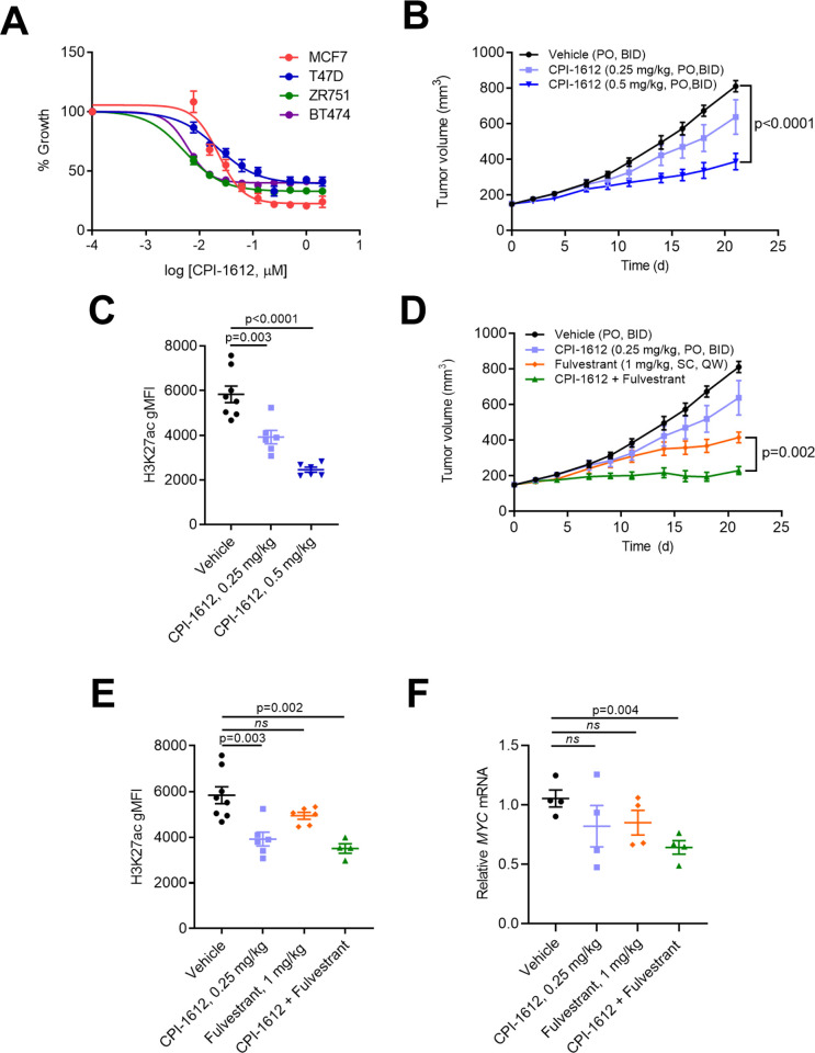
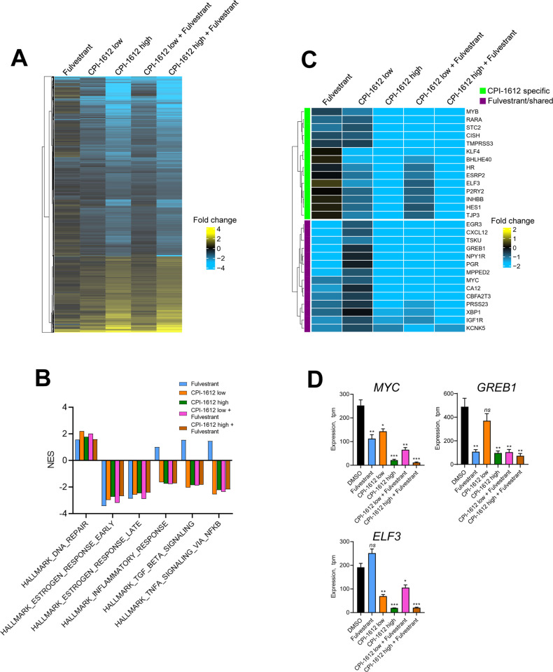
Therapeutic targeting of the estrogen receptor (ER) is a clinically validated approach for estrogen receptor positive breast cancer (ER+ BC), but sustained response is limited by acquired resistance. Targeting the transcriptional coactivators required for estrogen receptor activity represents an alternative approach that is not subject to the same limitations as targeting estrogen receptor itself. In this report we demonstrate that the acetyltransferase activity of coactivator paralogs CREBBP/EP300 represents a promising therapeutic target in ER+ BC. Using the potent and selective inhibitor CPI-1612, we show that CREBBP/EP300 acetyltransferase inhibition potently suppresses in vitro and in vivo growth of breast cancer cell line models and acts in a manner orthogonal to directly targeting ER. CREBBP/EP300 acetyltransferase inhibition suppresses ER-dependent transcription by targeting lineage-specific enhancers defined by the pioneer transcription factor FOXA1. These results validate CREBBP/EP300 acetyltransferase activity as a viable target for clinical development in ER+ breast cancer.


Conflict of interest statement

Authors are current or former employees and stockholders of Constellation Pharmaceuticals, a Morphosys company, which provided funding for this research. Patents have been filed around the chemical series that includes CPI-1612. This work does not relate to any marketed products or products in development. These disclosures do not alter our adherence to PLOS ONE policies on sharing data and materials.

Figures

Fig 1
Fig 1. CPI-1612 inhibits viability of ER+ breast cancer cell lines and ER signaling both in vitro and in vivo.
(A) In vitro activity of CPI-1612. ER+ breast cancer cell lines were treated with increasing doses of CPI-1612 and cell viability was measured using Cell Titer Glo after 4 days of treatment. Error bars represent standard deviation (n = 2). (B) In vivo activity of single agent CPI-1612. Female Balb/c nude mice were implanted subcutaneously with MCF7 cells (n = 8 for vehicle, n = 6 for others) and treated with the indicated doses of CPI-1612 (PO, BID) or an equal volume of vehicle (PO, BID). Tumor volumes were measured by caliper until study termination at 21 days. Data points represent mean and SEM at each timepoint. P-values were calculated using an unpaired student’s t-test relative to the vehicle arm; the p-value at study endpoint is shown (no data point in the 0.25 mg/kg arm reached statistical significance). (C) Pharmacodynamic readout of CPI-1612 activity. PBMCs were isolated from blood at study termination, fixed, and stained for FACS analysis. The level of H3K27ac was quantified using gMFI (geometric mean fluorescence intensity). Data are represented as mean ± SEM, and p-values were calculated using an unpaired student’s t-test. (D) Efficacy of CPI-1612 in combination with Fulvestrant. Mice were xenografted with MCF7 cells as in (B) and treated with CPI-1612 (0.25 mg/kg, PO, BID), Fulvestrant (1 mg/kg, SC, QW), CPI-1612 + Fulvestrant, or vehicle (n = 8 for vehicle, n = 6 for others). Data points represent the mean and SEM of surviving animals, and p-values were calculated at each timepoint using an unpaired student’s t-test. P-value for the difference between Fulvestrant and CPI-1612 + Fulvestrant at study endpoint is shown. (E) PD in PBMCs for study described in (D), as in (D). (F) Tumor PD as measured by gene expression changes. Total mRNA was isolated from tumors collected at study endpoint and used for q-RTPCR analysis. MYC expression normalized to ACTB was calculated relative to vehicle mean and is expressed as mean ± SEM for each arm. P-values were calculated by unpaired student’s t-test relative to vehicle.
Fig 2
Fig 2. CPI-1612 inhibits the ER transcriptional program.
(A) Bulk RNA-seq analysis of MCF7 cells. MCF7 cells were treated as indicated for 6 hours (n = 3 per treatment) followed by isolation of mRNA for RNA-sequencing analysis. Differential expression is indicated as log2 (fold-change) in normalized counts relative to the DMSO control. Genes shown were modulated at least 1.5-fold in at least one condition. Concentrations used were CPI-1612 low: 5 nM; CPI-1612 high: 50 nM; Fulvestrant: 100 nM. (B) CPI-1612 has a distinct transcriptional effect from Fulvestrant. Gene Set Enrichment Analysis (GSEA) was carried out using the MSigDB Hallmark genesets with the data described in (A). Normalized enrichment scores (NES) for selected genesets are shown. (C) CPI-1612 regulates the ER transcriptional network by impacting different genes than Fulvestrant. Subset of data in (A) showing selected genes in the HALLMARK_ESTROGEN_RESPONSE_EARLY geneset. Genes that are regulated by CPI-1612, Fulvestrant, or both are highlighted. (D) Example of differentially regulated genes. Expression values from RNA-seq are quantified as transcripts per million (tpm) and are plotted from the experiment described in (A). Values represent the mean and SEM for 3 replicates. P-values were calculated by unpaired student’s t-test (*: p<0.05; **:p<0.01;***:p<0.001; ns: not significant). P-values can be found in S3 Data.
Fig 3
Fig 3. CPI-1612 represses a subset of enhancers which are linked to differentially expressed genes.
(A) CPI-1612 treatment impacts chromatin accessibility and histone acetylation. MCF7 cells were treated for 6 hours with DMSO or CPI-1612 (50 nM), and samples were prepared for ATAC-seq or ChIP-seq with H3K27ac or H3K9ac antibodies. Waterfall plot of the log2 (fold-change) in signal intensity in open chromatin (ATAC-seq) peaks and H3K27ac peaks between DMSO and 50 nM CPI-1612 treatment. Blue: peaks with at least 2-fold decrease in signal; red: peaks with at least 2-fold increase in signal. (B) Global effects of CPI-1612 treatment. Top 30,000 peaks for each feature were ranked based on intensity for DMSO and CPI-1612 conditions. Graphs represent the sum of signal intensity across all peaks. (C) Fraction of peaks located in different genomic regions. Peaks from ATAC-seq or H3K27ac were assigned to the indicated genomic regions using HOMER. All: all peaks identified in DMSO and CPI-1612 conditions; differential: peaks that changed at least 2-fold upon treatment. (D) Genes mapped to differential ATAC-seq or H3K27ac peaks are likely to be downregulated. Genes were assigned to differential ATAC-seq or H3K27ac peaks, and differential expression data (as described in Fig 2) from DESeq2 were plotted. (E) Genes linked to differential features are enriched for ER targets. Genes from (D) were used for GSEA with Hallmark genesets. Top three signatures are shown with NES on the x-axis and adjusted P-value (Padj) next to bars. (F) Integrated gene expression and epigenomic features for the ELF3 locus. Top panel shows annotated genes, and purple boxes show annotated ELF3 enhancer elements. RNA-seq, H3K27ac ChIP-seq, and ATAC-seq tracks are plotted at the bottom, with H3K27ac and ATAC-seq peaks showing at least a 2-fold decrease marked with a *. The inset shows the differential ATAC-seq peaks in the ELF3 enhancer.
Fig 4
Fig 4. CPI-1612 targets FOXA1 binding sites that control luminal-specific gene sets in MCF7 cells and breast tumors.
(A) Differential ATAC-seq peaks are enriched for FOXA1 motifs. HOMER motif analysis was used to identify enrichment of transcription factor (TF) motifs in the ATAC-seq peaks that were downregulated at least 2-fold after CPI-1612 treatment, relative to the fraction of all ATAC-seq peaks with binding sites. (B) Downregulated genes and genes mapped to sites of reduced ATAC-seq signal are luminal-specific. GSEA was carried out on all genes (left) or genes mapped to ATAC-seq peaks that changed at least 2-fold (right) using either the Bas-ECJ or Lum(M)-ECJ genesets. NES and Padj values are indicated below the enrichment plots. (C) Differential ATAC-seq peaks are enriched for epigenomic features and TF binding. Published ChIP-seq data for the indicated features in MCF7 cells were plotted for the ATAC-seq peaks that were decreased at least 2-fold after CPI-1612 treatment. (D) Sites of differential ATAC-seq signal are more open in non-basal relative to basal breast tumors. Average ATAC-seq signal across all TCGA samples annotated as either basal or non-basal breast cancer was calculated for each of the differential ATAC-seq peaks described in Fig 3 and was compared to the difference in the average signal across a set of non-differential ATAC-seq peaks. P-values were calculated by unpaired student’s t-test with Welch’s correction on log-normalized read counts in each peak.

References

    1. Rosenberg PS, Barker KA, Anderson WF. Estrogen Receptor Status and the Future Burden of Invasive and In Situ Breast Cancers in the United States. J Natl Cancer Inst. 2015;107(9). doi: 10.1093/jnci/djv159 - DOI - PMC - PubMed
    1. Ali S, Buluwela L, Coombes RC. Antiestrogens and their therapeutic applications in breast cancer and other diseases. Annu Rev Med. 2011;62:217–32. doi: 10.1146/annurev-med-052209-100305 - DOI - PubMed
    1. Caswell-Jin JL, Plevritis SK, Tian L, Cadham CJ, Xu C, Stout NK, et al. Change in Survival in Metastatic Breast Cancer with Treatment Advances: Meta-Analysis and Systematic Review. JNCI Cancer Spectr. 2018;2(4):pky062. doi: 10.1093/jncics/pky062 - DOI - PMC - PubMed
    1. Osborne CK, Schiff R. Mechanisms of endocrine resistance in breast cancer. Annu Rev Med. 2011;62:233–47. doi: 10.1146/annurev-med-070909-182917 - DOI - PMC - PubMed
    1. Wang L, Tang Y, Cole PA, Marmorstein R. Structure and chemistry of the p300/CBP and Rtt109 histone acetyltransferases: implications for histone acetyltransferase evolution and function. Curr Opin Struct Biol. 2008;18(6):741–7. doi: 10.1016/j.sbi.2008.09.004 - DOI - PMC - PubMed