

Gastrointestinal biopsy in the horse: overview of collection, interpretation, and applications
- PMID: 35354416
- PMCID: PMC9254066
- DOI: 10.1177/10406387221085584
Gastrointestinal biopsy in the horse: overview of collection, interpretation, and applications
Abstract
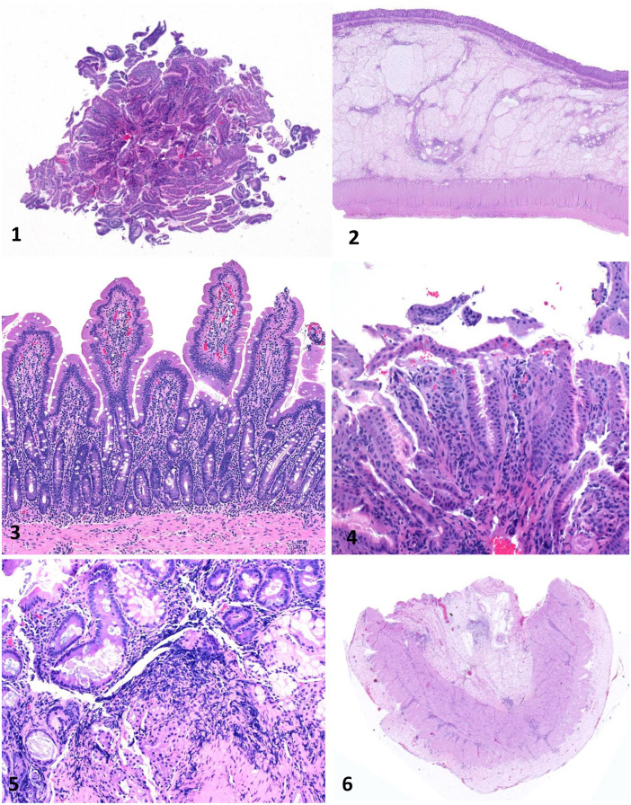
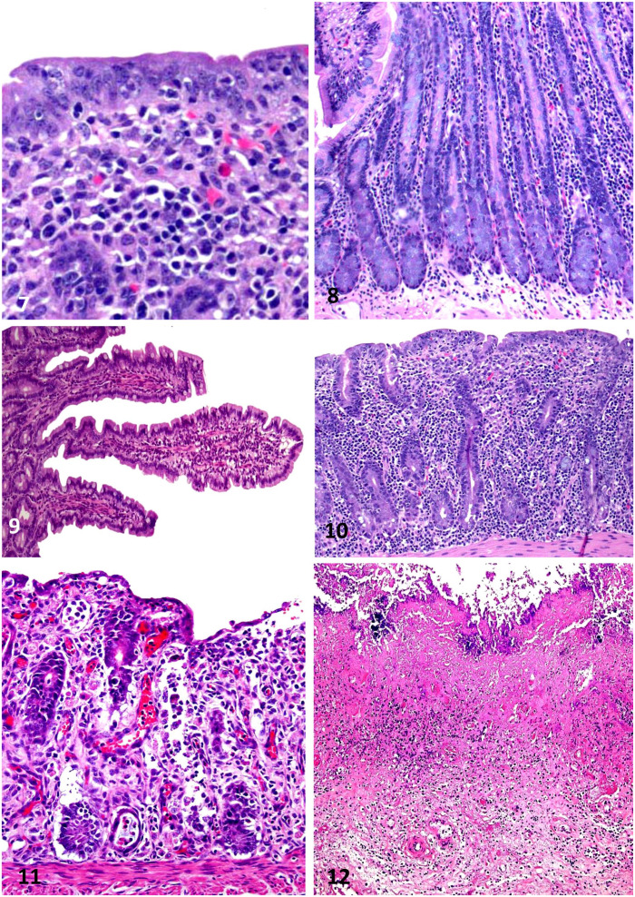
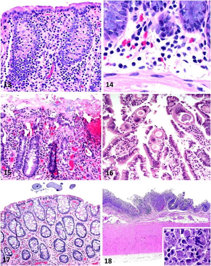
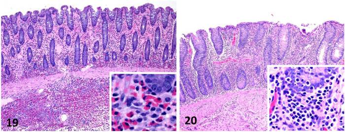
Evaluation of gastrointestinal (GI) biopsies is a multistep process that includes reviewing an appropriate history, determining sample quality, and evaluating histologic sections. Selected diagnostic parameters that, in combination with intestinal histopathology, can be useful to localize disease to the intestinal tract in the horse include hypoproteinemia and hypoalbuminemia, ultrasound evidence of increased thickness of the small intestinal wall, and alterations in glucose or D-xylose absorption tests. Biopsies may be acquired either endoscopically, or via laparoscopy or standing flank incisional approaches. GI sections should be evaluated using a systematic approach that includes both architectural changes and inflammatory cell infiltrates. Although strategies have been developed for assessment of GI biopsies from the dog and cat, a standardized approach to interpretation of the equine GI biopsy has yet to be developed. GI biopsies pose several challenges to the pathologist, especially for endoscopic biopsies in which the quality of the specimen and its orientation may vary greatly. Architectural changes are arguably the most critical changes to evaluate. In a horse with chronic GI inflammation, such as occurs in idiopathic inflammatory bowel disease (IBD), the cell types encountered frequently are macrophages, eosinophils, lymphocytes, and plasma cells. Increased numbers of these cell types are categorized loosely as mild, moderate, and severe. Specific forms of idiopathic IBD have been further classified by this infiltrate as granulomatous enteritis, eosinophilic enteritis, and lymphoplasmacytic enteritis; there is limited information on microscopic changes with each. Unfortunately, microscopic GI lesions are usually nonspecific, and determination of etiology requires further investigation.
Keywords: biopsy; gastrointestinal; horses; inflammatory bowel disease.
Conflict of interest statement
Figures
Similar articles
-
Idiopathic inflammatory bowel disease in dogs and cats: 84 cases (1987-1990).J Am Vet Med Assoc. 1992 Nov 15;201(10):1603-8. J Am Vet Med Assoc. 1992. PMID: 1289345
-
Clinicopathological and ultrasonographic features of cats with eosinophilic enteritis.J Feline Med Surg. 2014 Dec;16(12):950-6. doi: 10.1177/1098612X14525385. Epub 2014 Mar 3. J Feline Med Surg. 2014. PMID: 24591305 Free PMC article.
-
Immunohistochemical and morphometric analysis of intestinal full-thickness biopsy samples from cats with lymphoplasmacytic inflammatory bowel disease.J Comp Pathol. 2014 May;150(4):416-23. doi: 10.1016/j.jcpa.2014.01.002. Epub 2014 Jan 17. J Comp Pathol. 2014. PMID: 24612766
-
Chronic idiopathic inflammatory bowel diseases of the horse.J Vet Intern Med. 2000 May-Jun;14(3):258-65. doi: 10.1892/0891-6640(2000)014<0258:ciibdo>2.3.co;2. J Vet Intern Med. 2000. PMID: 10830538 Review.
-
Approach to Inflammatory Bowel Disease.Vet Clin North Am Equine Pract. 2024 Aug;40(2):287-306. doi: 10.1016/j.cveq.2024.04.004. Epub 2024 May 23. Vet Clin North Am Equine Pract. 2024. PMID: 38789349 Review.
Cited by
-
Tulathromycin and Diclazuril Lack Efficacy against Theileria haneyi, but Tulathromycin Is Not Associated with Adverse Clinical Effects in Six Treated Adult Horses.Pathogens. 2023 Mar 14;12(3):453. doi: 10.3390/pathogens12030453. Pathogens. 2023. PMID: 36986375 Free PMC article.
-
Ex vivo comparison of full-thickness biopsy techniques in the equine small intestine.Vet Surg. 2025 Jan;54(1):208-218. doi: 10.1111/vsu.14178. Epub 2024 Oct 15. Vet Surg. 2025. PMID: 39404177 Free PMC article.
-
Equine colic: A comprehensive overview of the sonographic evaluation, diagnostic criteria, and management of different categories.Open Vet J. 2025 Mar;15(3):1116-1139. doi: 10.5455/OVJ.2025.v15.i3.5. Epub 2025 Mar 31. Open Vet J. 2025. PMID: 40276205 Free PMC article. Review.
-
Histological Evaluation of Resected Tissue as a Predictor of Survival in Horses with Strangulating Small Intestinal Disease.Animals (Basel). 2023 Aug 26;13(17):2715. doi: 10.3390/ani13172715. Animals (Basel). 2023. PMID: 37684979 Free PMC article.
References
-
- Barr BS. Infiltrative intestinal disease. Vet Clin North Am Equine Pract 2006;22:e1–7. - PubMed
-
- Bianchi MV, et al.. Fatal parasite-induced enteritis and typhlocolitis in horses in Southern Brazil. Rev Bras Parasitol Vet 2019;28:443–450. - PubMed
-
- Camacho-Luna P, et al.. Advances in diagnostics and treatments in horses and foals with gastric and duodenal ulcers. Vet Clin North Am Equine Pract 2018;34:97–111. - PubMed
MeSH terms
LinkOut - more resources
Full Text Sources
Miscellaneous
