. 2022 Apr 15;208(8):2008-2018.
doi: 10.4049/jimmunol.2000945. Epub 2022 Mar 30.

IL-27 Enhances γδ T Cell-Mediated Innate Resistance to Primary Hookworm Infection in the Lungs

Affiliations

IL-27 Enhances γδ T Cell-Mediated Innate Resistance to Primary Hookworm Infection in the Lungs

Arjun Sharma et al. J Immunol. .

Abstract

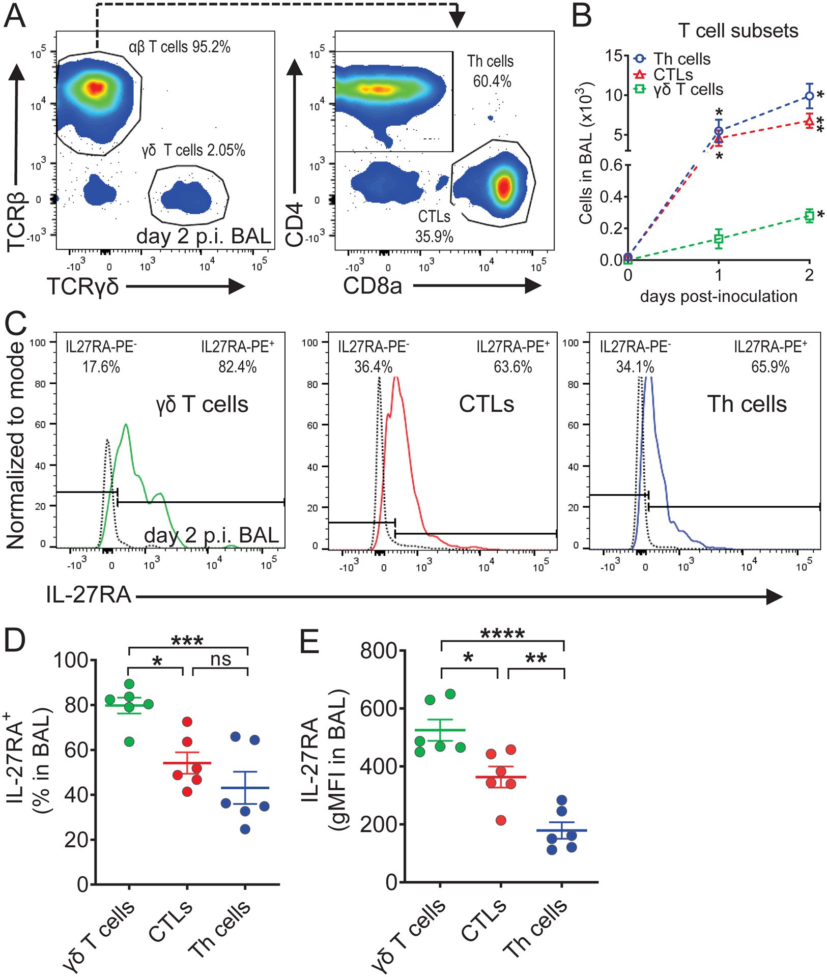
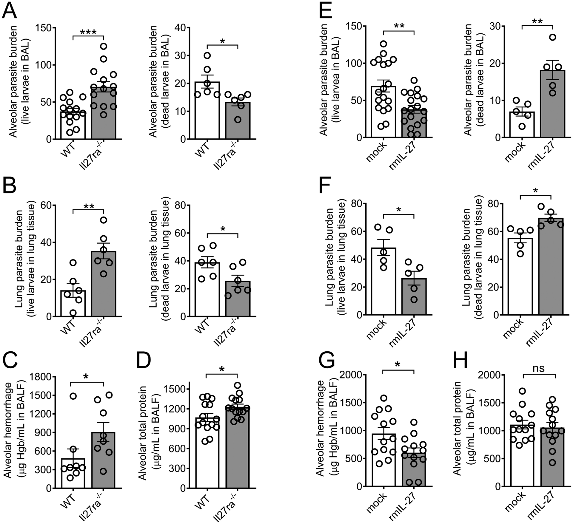
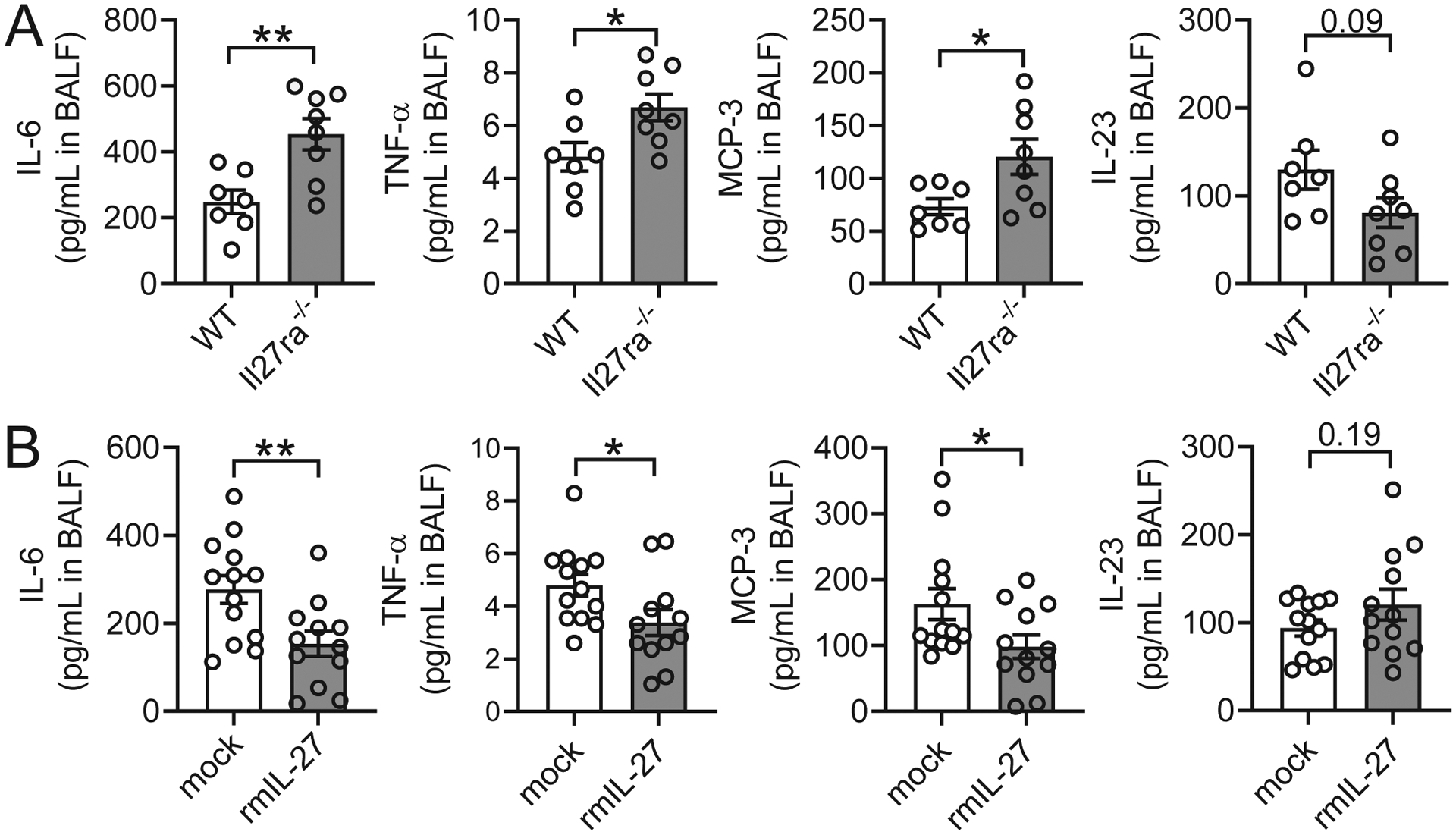
IL-27 is a heterodimeric IL-12 family cytokine formed by noncovalent association of the promiscuous EBI3 subunit and selective p28 subunit. IL-27 is produced by mononuclear phagocytes and unfolds pleiotropic immune-modulatory functions through ligation to IL-27 receptor α (IL-27RA). Although IL-27 is known to contribute to immunity and to limit inflammation after various infections, its relevance for host defense against multicellular parasites is still poorly defined. Here, we investigated the role of IL-27 during infection with the soil-transmitted hookworm, Nippostrongylus brasiliensis, in its early host intrapulmonary life cycle. IL-27(p28) was detectable in bronchoalveolar lavage fluid of C57BL/6J wild-type mice on day 1 after s.c. inoculation. IL-27RA expression was most abundant on lung-invading γδ T cells. Il27ra-/- mice showed increased lung parasite burden together with aggravated pulmonary hemorrhage and higher alveolar total protein leakage as a surrogate for epithelial-vascular barrier disruption. Conversely, injections of recombinant mouse (rm)IL-27 into wild-type mice reduced lung injury and parasite burden. In multiplex screens, higher airway accumulations of IL-6, TNF-α, and MCP-3 (CCL7) were observed in Il27ra-/- mice, whereas rmIL-27 treatment showed a reciprocal effect. Importantly, γδ T cell numbers in airways were enhanced by endogenous or administered IL-27. Further analysis revealed a direct antihelminthic function of IL-27 on γδ T cells as adoptive intratracheal transfer of rmIL-27-treated γδ T cells during primary N. brasiliensis lung infection conferred protection in mice. In summary, this report demonstrates protective functions of IL-27 to control the early lung larval stage of hookworm infection.


Conflict of interest statement

Disclosures

The authors have no financial conflicts of interest.

Figures

FIGURE 1.
FIGURE 1.
N. brasiliensis infection promotes IL-27(p28) release and IL-27RA-expressing lymphocytes in the lungs. C57BL/6J wild type (WT) mice were infected s.c. with third stage larvae (L3) of N. brasiliensis (n=500/mouse) or received a mock PBS injection as controls. The inflammatory cytokines or cells were collected by broncho-alveolar lavage (BAL). (A) IL-27(p28) was quantified in BAL fluid by ELISA at the indicated time points (n=3–5 mice/group). (B) Representative flow cytometry gating of CD19 versus CD3 pre-gated on CD45+Ly6G single cell lymphocytes (left panel), and NK1.1 versus CD3 gated on CD19CD3 innate lymphocytes (right panel), on day 2 post-inoculation (p.i.). Frequencies (%) of parent populations are indicated next to each gate. (C) Absolute numbers of lymphocyte populations of CD19CD3+ T cells, CD19+CD3 B cells, and CD19CD3NK1.1+ NK cells on days 0 (PBS-inoculated/uninfected), 1, and 2 p.i. (n=6–9 mice/group). (D) Representative histograms of IL-27RA expression on T cells (left panel), NK cells (middle panel), and B cells (right panel) on day 2 p.i. The dotted black line indicates isotype-FMO control. The frequencies of IL-27RA and IL-27RA+ cells are indicated in the upper left and right corners, respectively. (E) Frequencies of IL-27RA+ T cells, NK cells, and B cells on days 1 and 2 p.i. (n=8–9 mice/group from 2 pooled experiments). (F) IL-27RA presence expressed as geometric mean fluorescence intensity (gMFI) on these cells at these time points (n=6 mice/group). (G) Representative histogram of IL-27RA expression on T cells on days 1 and 2 p.i. The dotted black line indicates isotype-FMO control (Ctrl). Each circle indicates an individual mouse (A, E, F). Data are shown as mean ± SEM (A, C, E, F). Data were analyzed by Student’s t-test comparing day 0 vs. day 1 and day 0 vs. day 2 for each cell type (C), one-way ANOVA (A), or two-way ANOVA (E, F), * P<0.05, ** P<0.01, *** P<0.001, **** P<0.0001, ns: not significant.
FIGURE 2.
FIGURE 2.
IL-27RA expression is highest on γδ T cells among broncho-alveolar lymphocytes during N. brasiliensis infection. C57BL/6J (WT) mice were infected s.c. with third stage larvae (L3) of N. brasiliensis (n=500/mouse) or received a mock PBS injection as controls. Cells were collected by BAL. (A) Representative flow cytometry gating of TCRβ chain versus TCRγδ chain pre-gated on CD3+ single cell lymphocytes (left panel), and CD4 versus CD8a on αβ T cells (right panel), on day 2 p.i. Frequencies (%) of parent populations are indicated next to each gate. (B) Absolute numbers of CD4+CD8 T helper (Th) cells, CD4CD8+ cytotoxic T lymphocytes (CTLs), and TCRβTCRγδ+ γδ T cells on day 0 (PBS-inoculated/uninfected), day 1 (n=2 mice/group), and day 2 (n=6 mice/group) p.i. (C) Representative histograms of IL-27RA expression on γδ T cells (left panel), CTLs (middle panel), and Th cells (right panel). The dotted black line indicates IL-27RA staining in Il27ra−/− mice as a negative control. The frequencies of IL-27RA and IL-27RA+ cells are indicated in the upper left and right corners, respectively. (D) Frequencies of IL-27RA+ γδ T cells, CTLs, and Th cells on day 2 p.i. (n=6 mice/group). (E) IL-27RA abundance (gMFI) on γδ T cells, CTLs, and Th cells on day 2 p.i. (n=6 mice/group). Each circle indicates an individual mouse (D, E). Data are shown as mean ± SEM (B, D, E). Data were analyzed by Student’s t-test comparing day 0 vs. day 1 and day 0 vs. day 2 for each cell type (B), or one-way ANOVA (D, E), * P<0.05, ** P<0.01, *** P<0.001, **** P<0.0001, ns: not significant.
FIGURE 3.
FIGURE 3.
IL-27 enhances innate resistance to primary N. brasiliensis infection in the lungs. (A-D) In vivo comparison of the susceptibility of Il27ra−/− mice and WT control (C57BL/6NJ) mice to primary N. brasiliensis infection (n=500 L3/mouse, s.c.) in the lungs on day 2 p.i. (A) Alveolar live and dead parasite burden in BAL (n=6–14 mice/group) and (B) parasite burden within lung tissue consisting of live and dead larvae (n=6 mice/group). (C) Alveolar hemorrhage (n=8 mice/group, and (D) alveolar total protein in BALF (n=14 mice/group). (E-H) In vivo comparison of the susceptibility of C57BL/6J mice administered rmIL-27 (100 ng/mouse) i.p. on days 0 and 1 p.i.) or mock control (0.1% BSA in PBS i.p.) to primary N. brasiliensis infection in the lungs on day 2 p.i. (E) Alveolar parasite burden in BAL and (F) lung parasite burden within tissue consisting of live and dead larvae (n=18 or n=5 mice/group). (G) Alveolar hemorrhage and (H) alveolar total protein in BALF (n=13 mice/group from 2 pooled experiments). Each circle indicates an individual mouse. Frames A, D, G-H are pooled from two independent experiments and data are shown as mean ± SEM and were analyzed by Student’s t-test, * P<0.05, ** P<0.01, *** P<0.001, ns: not significant.
FIGURE 4.
FIGURE 4.
IL-27 regulates inflammatory mediators during primary N. brasiliensis infection of the lungs. (A) In vivo comparison of selected BALF cytokines and chemokines from Il27ra−/− mice and WT control (C57BL/6NJ) mice during primary N. brasiliensis infection (n=500 L3/mouse, s.c.) on day 2 p.i. (n=7–8 mice/group). (B) In vivo comparison of selected BALF cytokines and chemokines in WT (C57BL/6J) mice administered rmIL-27 (100 ng/mouse i.p on days 0 and 1 p.i.) or mock control (0.1% BSA in PBS i.p.) during primary N. brasiliensis infection on day 2 p.i. (n=12–13 mice/group from 2 pooled experiments). All data were obtained by bead-based multiplex assay (Luminex-200). Each circle indicates an individual mouse. Data are shown as mean ± SEM and were analyzed by Student’s t-test, * P<0.05, ** P<0.01.
FIGURE 5.
FIGURE 5.
Il27ra−/− mice have decreased presence of γδ T cells in the alveolar space during N. brasiliensis infection. (A) Representative flow cytometry gating of TCRβTCRγδ+ γδ T cells (top panels), CD4+CD8 Th cells, and CD4CD8+ CTLs (bottom panels), from WT control (C57BL/6NJ) (left panels) and Il27ra−/− mice (right panels) in BAL during N. brasiliensis infection (n=500 L3/mouse, s.c.) on day 2 p.i. Frequencies (%) of parent populations are indicated next to each gate. Cells were pre-gated on live singlet lymphocytes. (B) Frequencies and (C) absolute numbers of these CD4+CD8 Th cells, CD4CD8+ CTLs, and TCRβTCRγδ+ γδ T cells in BAL of WT (n=5 mice/group; one mouse was removed due to extremely low infection, as determined by negligible hemorrhage) and Il27ra−/− (n=6 mice/group) mice on day 2 p.i. Each circle indicates an individual mouse (B, C). Data are shown as mean ± SEM (B, C) and were analyzed by Student’s t-test (B,C), * P<0.05, ns: not significant.
FIGURE 6.
FIGURE 6.
IL-27 enhances γδ T cell-mediated innate resistance to primary N. brasiliensis infection in the lungs. (A-B) C57BL/6J (WT) mice were administered rmIL-27 (100 ng/mouse i.p. on days 0 and 1 p.i.) or mock control (0.1% BSA in PBS i.p.) during N. brasiliensis infection (n=500 L3/mouse, s.c.). γδ T cells were analyzed on day 2 p.i in BAL. (A) Representative flow cytometry plots showing frequencies and (B) absolute numbers of CD3+ TCRγδ+ γδ T cells in the alveolar spaces of rm-IL27-treated mice (n=5 mice/group). (C) Workflow demonstrating γδ T cell adoptive transfer approach. γδ T cells (CD11bCD45RCD3+TCRγδ+) were positively selected from the spleens of naïve WT mice using magnetic beads (purity of TCRγδ+ fraction ~95%), treated for 30 min at 37°C in vitro with mock (PBS) or rmIL-27 (100 ng/ml), washed thoroughly, and injected intratracheally into recipient mice 12 hours after inoculation with N. brasiliensis. (D) Alveolar parasite burden by BAL and (E) lung parasite burden within lung tissue consisting of live and dead larvae on day 2 p.i. following the adoptive transfer (n=6–7 mice/group). Each circle indicates an individual mouse (B, D, E). Data are shown as mean ± SEM (B, D, E) and were analyzed by Student’s t-test, * P<0.05, ** P<0.01, *** P<0.001.

Similar articles

Cited by

References

    1. Loukas A, Hotez PJ, Diemert D, Yazdanbakhsh M, McCarthy JS, Correa-Oliveira R, Croese J, and Bethony JM. 2016. Hookworm infection. Nat Rev Dis Primers 2: 16088. - PubMed
    1. Pullan RL, Smith JL, Jasrasaria R, and Brooker SJ. 2014. Global numbers of infection and disease burden of soil transmitted helminth infections in 2010. Parasit Vectors 7: 37. - PMC - PubMed
    1. Herricks JR, Hotez PJ, Wanga V, Coffeng LE, Haagsma JA, Basanez MG, Buckle G, Budke CM, Carabin H, Fevre EM, Furst T, Halasa YA, King CH, Murdoch ME, Ramaiah KD, Shepard DS, Stolk WA, Undurraga EA, Stanaway JD, Naghavi M, and Murray CJL. 2017. The global burden of disease study 2013: What does it mean for the NTDs? PLoS Negl Trop Dis 11: e0005424. - PMC - PubMed
    1. Brooker S, Hotez PJ, and Bundy DA. 2008. Hookworm-related anaemia among pregnant women: a systematic review. PLoS Negl Trop Dis 2: e291. - PMC - PubMed
    1. Smith JL, and Brooker S. 2010. Impact of hookworm infection and deworming on anaemia in non-pregnant populations: a systematic review. Trop Med Int Health 15: 776–795. - PMC - PubMed