

Case Reports
doi: 10.1136/svn-2021-001407.
Epub 2022 Mar 30.
Retrograde recanalisation for vertebral artery stump syndrome: a case report
Affiliations
- PMID: 35354663
- PMCID: PMC9614125
- DOI: 10.1136/svn-2021-001407
Item in Clipboard
Case Reports
Retrograde recanalisation for vertebral artery stump syndrome: a case report
Stroke Vasc Neurol.
2022 Oct.
Abstract
Vertebral artery stump syndrome (VASS) is a rare disease associated with a posterior circulation stroke after vertebral artery origin occlusion. We have herein presented a case of VASS that was effectively treated with endovascular intervention using retrograde recanalisation and the mechanism of VASS in our patient was thought to be a thrombus formed by stagnating flow.
Keywords: atherosclerosis; stents; stroke.
© Author(s) (or their employer(s)) 2022. Re-use permitted under CC BY-NC. No commercial re-use. See rights and permissions. Published by BMJ.
Conflict of interest statement
Competing interests: None declared.
Figures
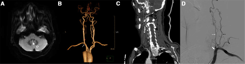
(A) Diffusion-weighted imaging at admission showed an acute ischaemic stroke in the left cerebellar hemisphere and vermis. (B) CT angiography revealed an occlusion at the origin of the left vertebral artery (VA) (white arrow). (C) CT angiography revealed atherosclerotic calcification of left V4 segment (white arrow). (D) Angiography detected total occlusion at the origin of left VA (white arrowhead), with distal antegrade collateral flow via the ascending cervical artery originating from the thyrocervical trunk (white arrow) and the deep cervical artery (black arrow) at the C3 level (oblique projection).
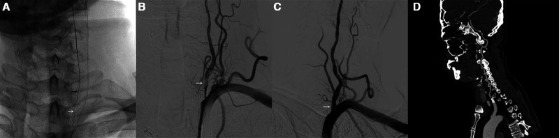
(A) Synchro-2 microwire and SL-10 microcatheter passed through the ascending cervical artery and reached the left vertebral ostium and then to the left subclavian artery (black arrow), using Synchro-2 wire as a marker, a PT-2 microwire (0.014 in×300 cm) successfully found the true lumen of the left VA (white arrow). (B) Filling defect found at the origin of left VA (white arrow). (C) Angiography after the bare-metal stent (white arrow) planted. (D) Follow-up at 6 months, CT angiography (CTA) showed no restenosis in the stent.
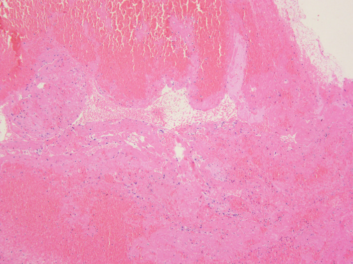
Histopathology of the removed thrombus which contained neutrophils, platelets and red blood cells within a dense laminar fibrin network.
References
Publication types
MeSH terms
Supplementary concepts
LinkOut - more resources
Full Text Sources
Medical