Radiation safety for pain physicians: principles and recommendations
- PMID: 35354676
- PMCID: PMC8977205
- DOI: 10.3344/kjp.2022.35.2.129
Radiation safety for pain physicians: principles and recommendations
Abstract
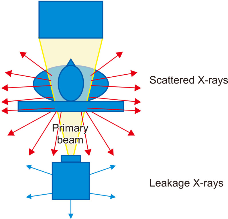
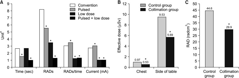
C-arm fluoroscopy is a useful tool for interventional pain management. However, with the increasing use of C-arm fluoroscopy, the risk of accumulated radiation exposure is a significant concern for pain physicians. Therefore, efforts are needed to reduce radiation exposure. There are three types of radiation exposure sources: (1) the primary X-ray beam, (2) scattered radiation, and (3) leakage from the X-ray tube. The major radiation exposure risk for most medical staff members is scattered radiation, the amount of which is affected by many factors. Pain physicians can reduce their radiation exposure by use of several effective methods, which utilize the following main principles: reducing the exposure time, increasing the distance from the radiation source, and radiation shielding. Some methods reduce not only the pain physician's but also the patient's radiation exposure. Taking images with collimation and minimal use of magnification are ways to reduce the intensity of the primary X-ray beam and the amount of scattered radiation. It is also important to carefully select the C-arm fluoroscopy mode, such as pulsed mode or low-dose mode, for ensuring the physician's and patient's radiation safety. Pain physicians should practice these principles and also be aware of the annual permissible radiation dose as well as checking their radiation exposure. This article aimed to review the literature on radiation safety in relation to C-arm fluoroscopy and provide recommendations to pain physicians during C-arm fluoroscopy-guided interventional pain management.
Keywords: Fluoroscopy; Interventional; Ionizing; Pain; Procedural; Radiation; Radiation Dosage; Radiation Effects; Radiation Exposure; Radiography; Radiology; Safety; Scattering; X-Rays..
Conflict of interest statement
No potential conflict of interest relevant to this article was reported.
Figures
Similar articles
-
Ionizing Radiation Dose Exposure to the Ocular Region of Pain Physicians During C-arm Guided Pain Interventions.Pain Physician. 2018 Sep;21(5):E523-E532. Pain Physician. 2018. PMID: 30282400
-
Radiation Exposure in Interventional Pain Management Physicians: A Systematic Review of the Current Literature.Pain Physician. 2024 Jan;27(1):E17-E35. Pain Physician. 2024. PMID: 38285025
-
Directional vector visualization of scattered rays in mobile c-arm fluoroscopy.Radiol Phys Technol. 2024 Mar;17(1):288-296. doi: 10.1007/s12194-024-00779-w. Epub 2024 Feb 5. Radiol Phys Technol. 2024. PMID: 38316688
-
Radiation safety: a focus on lead aprons and thyroid shields in interventional pain management.Korean J Pain. 2018 Oct;31(4):244-252. doi: 10.3344/kjp.2018.31.4.244. Epub 2018 Oct 1. Korean J Pain. 2018. PMID: 30310549 Free PMC article. Review.
-
Radiation risk management during fluoroscopy for interventional pain medicine physicians.Curr Pain Headache Rep. 2004 Feb;8(1):49-55. doi: 10.1007/s11916-004-0040-x. Curr Pain Headache Rep. 2004. PMID: 14731383
Cited by
-
Influence of Obesity, Race and Gender on Radiation Exposure for Epidural Procedures.Curr Pain Headache Rep. 2025 Jan 6;29(1):12. doi: 10.1007/s11916-024-01327-2. Curr Pain Headache Rep. 2025. PMID: 39760932 Review.
-
Augmented Reality-Assisted Navigation System for Transforaminal Epidural Injection.J Pain Res. 2023 Mar 17;16:921-931. doi: 10.2147/JPR.S400955. eCollection 2023. J Pain Res. 2023. PMID: 36960464 Free PMC article.
-
Comparison of international medical costs for interventional pain treatment: a focus on Korea and Japan.Korean J Pain. 2024 Jan 1;37(1):51-58. doi: 10.3344/kjp.23254. Epub 2023 Dec 11. Korean J Pain. 2024. PMID: 38072796 Free PMC article.
-
Clinical Application of an Augmented Reality Navigation System for Transforaminal Epidural Injection: A Randomized Controlled Trial.J Clin Med. 2024 Mar 29;13(7):1992. doi: 10.3390/jcm13071992. J Clin Med. 2024. PMID: 38610758 Free PMC article.
-
Evaluation of a first of a kind robotic radiation protection technology to reduce scatter exposure during diagnostic procedures and percutaneous coronary interventions.Am Heart J Plus. 2025 Feb 21;52:100512. doi: 10.1016/j.ahjo.2025.100512. eCollection 2025 Apr. Am Heart J Plus. 2025. PMID: 40093308 Free PMC article.
References
-
- Milacic S. Risk of occupational radiation-induced cataract in medical workers. Med Lav. 2009;100:178–86. - PubMed
Publication types
LinkOut - more resources
Full Text Sources