. 2022 Mar 30;12(1):5375.
doi: 10.1038/s41598-022-09306-6.

Human liver organoid derived intra-hepatic bile duct cells support SARS-CoV-2 infection and replication

Affiliations

Human liver organoid derived intra-hepatic bile duct cells support SARS-CoV-2 infection and replication

Vincent Chi-Hang Lui et al. Sci Rep. .

Abstract

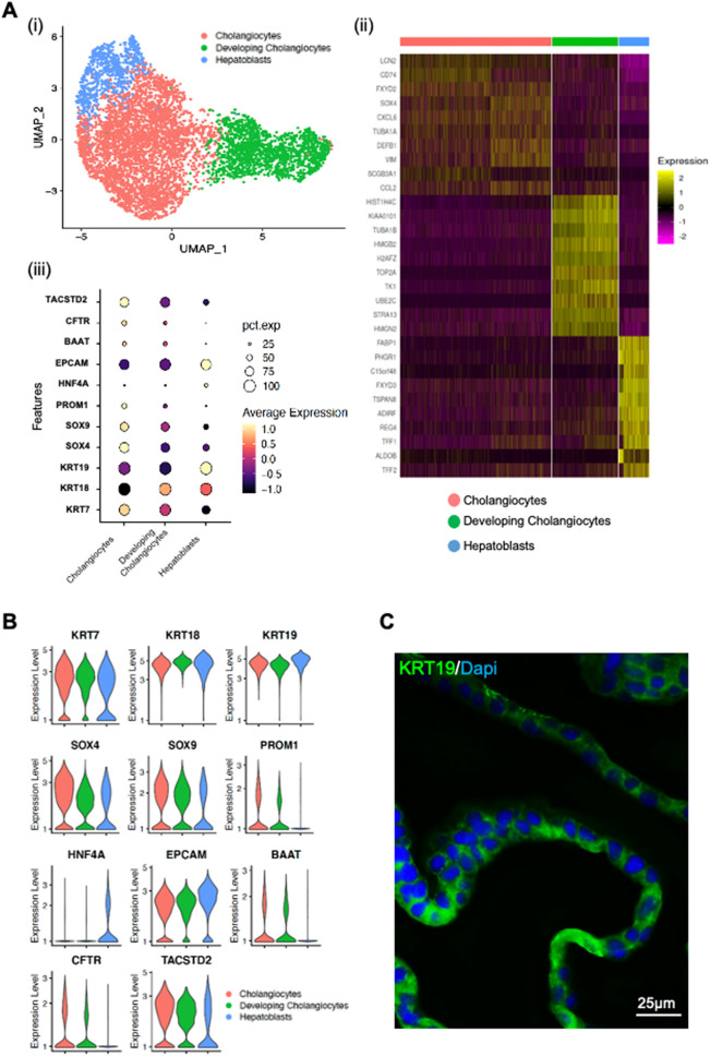
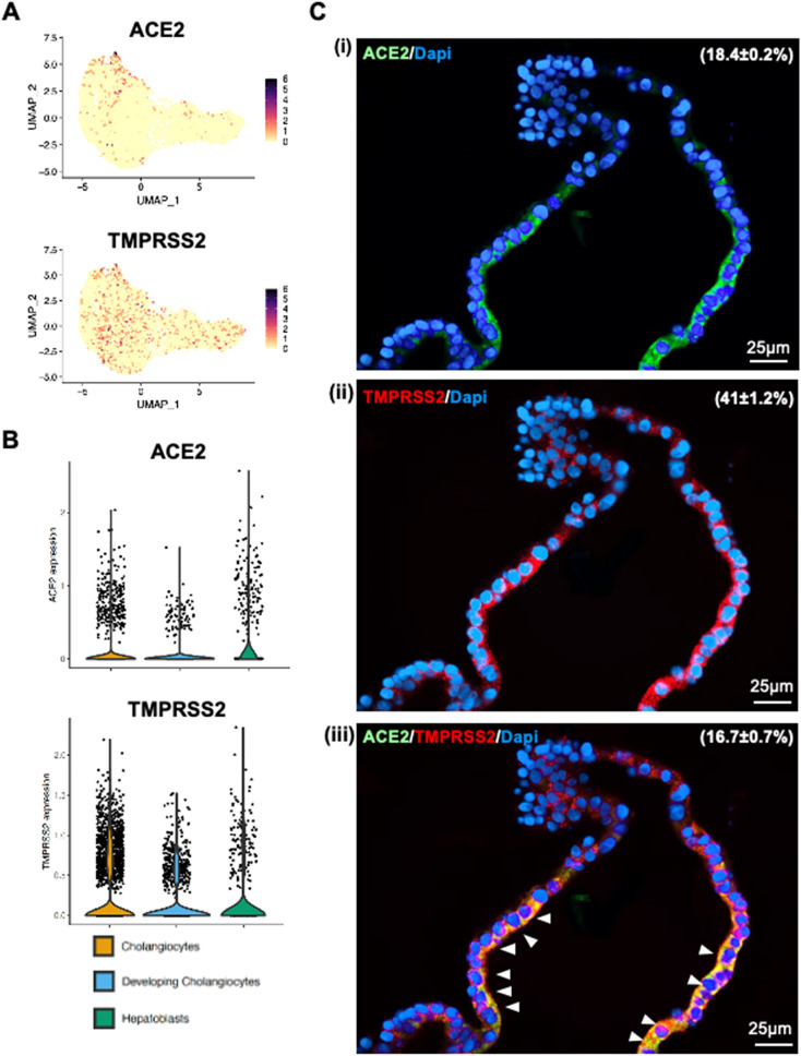
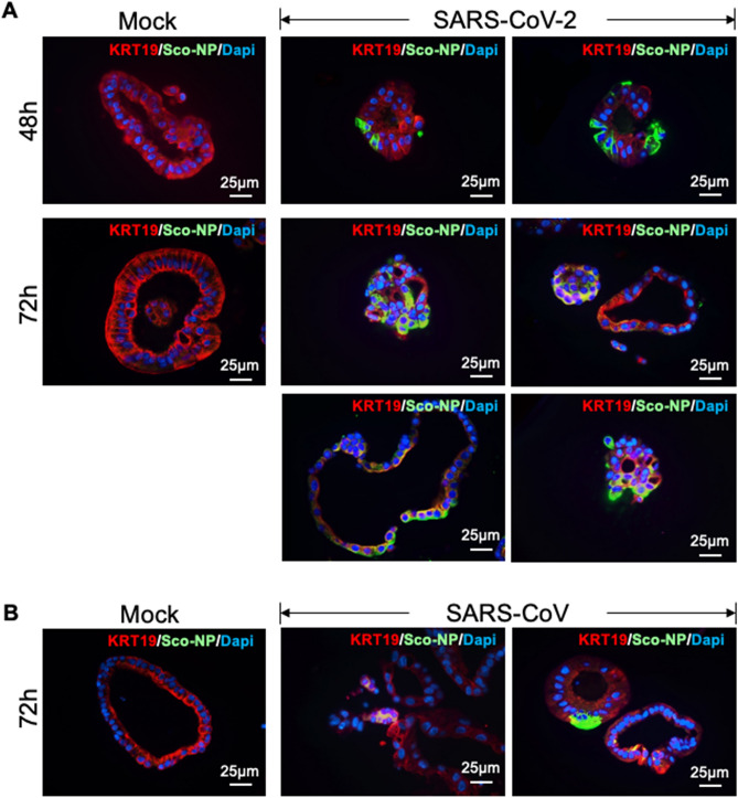
Although the main route of infection for severe acute respiratory syndrome coronavirus 2 (SARS-CoV-2) is the respiratory tract, liver injury is also commonly seen in many patients, as evidenced by deranged parenchymal liver enzymes. Furthermore, the severity of liver damage has been shown to correlate with higher mortality. Overall, the mechanism behind the liver injury remains unclear. We showed in this study that intra-hepatic bile duct cells could be grown using a human liver organoid platform. The cholangiocytes were not only susceptible to SARS-CoV-2 infection, they also supported efficient viral replication. We also showed that SARS-CoV-2 replication was much higher than SARS-CoV. Our findings suggested direct cytopathic viral damage being a mechanism for SARS-CoV-2 liver injury.


Conflict of interest statement

All authors declare no support from any organization for the submitted work; no financial relationships with any organizations that might have an interest in the submitted work in the previous three years; no other relationships or activities that could appear to have influenced the submitted work.

Figures

Figure 1
Figure 1
Human liver organoid cells expressed markers of cholangiocyte and hepatoblast. Characterisation of human liver organoids. (A(i)) UMAP showing the assigned identity for each cluster (3 clusters) in 10X Genomics single-cell RNA sequencing analysis of human control liver organoids. (A(ii)) Heatmap of top 10 markers expressed for each clusters in HB control organoids at p < 0.05. (A(iii)) Dot plot showing expression of canonical markers used for identification of clusters including cytokeratin-19 (KRT19) and epithelial cellular adhesion molecule (EPCAM). (B) Violin plots showing levels of expression of canonical markers for cholangiocytes and hepatoblast used for classification of cell types in control liver organoids. (C) Immunofluorescence staining for KRT19 of human control liver organoids.
Figure 2
Figure 2
Human liver organoid cells expressed ACE2 and TMPRS2. (A) and (B) Feature plot and violin plot displaying single cell expression distributions for features (ACE2, TMPRSS2 and TMPRSS4) in each cluster of cells from liver organoids. (C) Co-immunofluorescence staining of human liver organoids for ACE2 and TMPRSS2. Parenthesis indicated the percentages of organoid cells that expressed ACE2 (i), TMPRSS2 (ii) and ACE2 plus TMPRSS2 (iii). Arrowheads, organoid cells that co-expressed ACE2 and TMPRSS2.
Figure 3
Figure 3
Human liver organoids are susceptible to infection by SARS-CoV-2 and other coronaviruses. (A) Immunofluorescence staining for SARS-CoV nucleocapsid protein (Sco-Np) of mock and SARS-CoV-2 infected organoids at 48 h and 72 h post-infection (4 organoids shown). (B) Immunofluorescence staining for SARS-CoV nucleocapsid protein (Sco-Np) of mock and SARS-CoV infected organoids at 72 h post-infection.
Figure 4
Figure 4
Viral infection and replication kinetics of SARS-CoV-2 and SARS-CoV in human liver organoids. Bar chart showing viral titres in culture supernatants of human liver organoids infected with SARS-CoV-2 and SARS-CoV viruses. The organoids were infected at MOI 0.1 at 37 °C. Culture supernatants were harvested at the indicted times and virus titres were measured by TCID50 assay. Bar-charts show the mean virus titre ± standard deviation (SD). The horizontal dotted line denotes the limit of detection in the TCID50 assay. Statistical significances compared among viruses were calculated using two-way ANOVA with Tukey’s multiple comparisons test. ***p < 0·001. (C) Bar-charts showing the area under the curve (AUC) derived from the viral titres from 24 to 72 h. Data are presented as mean ± standard deviation (SD). Statistical significances compared among viruses were calculated using one-way ANOVA with Tukey’s multiple comparisons test. ***p < 0·001.

References

    1. From Wolrd Health Organisation. https://covid19.who.int (accessed 3rd February 2022)
    1. Wu F, Zhao S, Yu B, Chen YM, Wang W, Song ZG, et al. A new coronavirus associated with human respiratory disease in China. Nature. 2020;579:265–269. doi: 10.1038/s41586-020-2008-3. - DOI - PMC - PubMed
    1. Hoffmann M, Kleine-Weber H, Schroeder S, Kruger N, Herrler T, Erichsen S, et al. SARS-CoV-2 cell entry depends on ACE2 and TMPRSS2 and is blocked by a clinically proven protease inhibitor. Cell. 2020;181:271–280. doi: 10.1016/j.cell.2020.02.052. - DOI - PMC - PubMed
    1. Stopsack KH, Mucci LA, Antonarakis ES, Nelson PS, Kantoff PW. TMPRSS2 and COVID-19: serendipity or opportunity for intervention? Cancer Discov. 2020;10:779–782. doi: 10.1158/2159-8290.CD-20-0451. - DOI - PMC - PubMed
    1. Wu Z, McGoogan JM. Characteristics of and Important lessons from the coronavirus disease 2019 (COVID-19) outbreak in china: summary of a report of 72314 cases from the chinese center for disease control and prevention. JAMA. 2020;323:1239–1242. doi: 10.1001/jama.2020.2648. - DOI - PubMed

Publication types