. 2022 Mar 30;12(1):5350.
doi: 10.1038/s41598-022-09383-7.

Influence of air pollutants on circulating inflammatory cells and microRNA expression in acute myocardial infarction

Affiliations

Influence of air pollutants on circulating inflammatory cells and microRNA expression in acute myocardial infarction

Alberto Cecconi et al. Sci Rep. .

Abstract

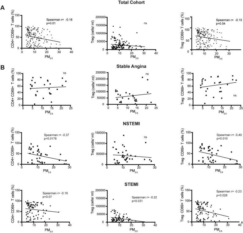
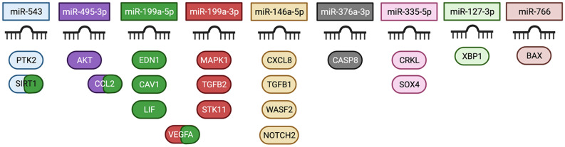
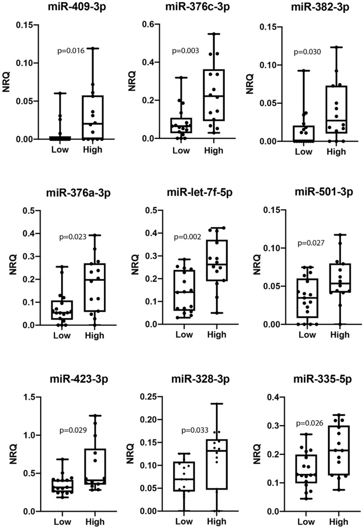
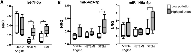
Air pollutants increase the risk and mortality of myocardial infarction (MI). The aim of this study was to assess the inflammatory changes in circulating immune cells and microRNAs in MIs related to short-term exposure to air pollutants. We studied 192 patients with acute coronary syndromes and 57 controls with stable angina. For each patient, air pollution exposure in the 24-h before admission, was collected. All patients underwent systematic circulating inflammatory cell analyses. According to PM2.5 exposure, 31 patients were selected for microRNA analyses. STEMI patients exposed to PM2.5 showed a reduction of CD4+ regulatory T cells. Furthermore, in STEMI patients the exposure to PM2.5 was associated with an increase of miR-146a-5p and miR-423-3p. In STEMI and NSTEMI patients PM2.5 exposure was associated with an increase of miR-let-7f-5p. STEMI related to PM2.5 short-term exposure is associated with changes involving regulatory T cells, miR-146a-5p and miR-423-3p.


Conflict of interest statement

The authors declare no competing interests.

Figures

Figure 1
Figure 1
Correlation map of air pollutants. **Correlation is significant at the 0.01 level. *Correlation is significant at the 0.05 level. X no significant correlation.
Figure 2
Figure 2
Correlation of PM2.5 with CD69 + CD4 T cells and Treg cells. Scatter plots are shown. (A) Association of PM2.5 exposure with CD4 + T cell subsets in the whole cohort. (B) Association of PM2.5 exposure with CD4 + T cell subsets in stable angina, NSTEMI and STEMI patients. Correlation was assessed by Spearman test.
Figure 3
Figure 3
Correlations of CO and SO2 exposure with circulatory immune cells. Scatter plots of significant correlations are shown. Correlation was assessed by Spearman test. (A) CO exposure had a positive correlation with the percentage of CD4+T cells as well as with IL22- and IL-17-producers CD4+ T cells. (B) Negative correlation of CO exposure with CD4+CD69+ lymphocytes and CD69+ Treg cells. (C) SO2 exposure was associated with a decrease of total leucocytes count.
Figure 4
Figure 4
Target genes of differentially expressed genes in CAD patients exposed to high levels of PM2.5. Functional miRNA targets associated to both Cardiovascular System and Immune System are shown. Different colors are used to indicate the targets that are regulated by each miRNA. From all genes identified in miRTarBase as targets of microRNAs listed in Table 2, only those with functioonal support were selected to perform the enrichment analysis. Image created with BioRender.com.
Figure 5
Figure 5
Differential expression of miRNAs in plasma samples from CAD patients exposed to low and high levels of PM2.5. Box and whiskers Min to Max plots showing plasma levels of microRNAs from CAD patients (n = 31) exposed to low or high levels of PM2.5. Including all clinical presentations in the analysis, high PM2.5 short-term exposure was associated with (A) increased and (B) decreased miRNA expression. Differences were analyzed using Mann–Whitney U test.
Figure 6
Figure 6
Circulating levels of miR-let-7f-5p, miR-423-3p and miR-146a-5p are increased in acute myocardial patients exposed to high levels of PM2.5. Box and whiskers Min to Max plots showing the expression of (A) miR-let-7f-5p and (B) miR-423-3p and miR-146a-5p in plasma samples from stable angina patients (n = 8), NSTEMI patients (n = 9) and STEMI patients (n = 14) exposed to low levels (empty boxes) or high levels (grey boxes) of PM2.5. Differences were analyzed using Mann–Whitney U test, *p < 0.01.

Similar articles

Cited by

References

    1. Lelieveld J, Klingmüller K, Pozzer A, Pöschl U, Fnais M, Daiber A, et al. Cardiovascular disease burden from ambient air pollution in Europe reassessed using novel hazard ratio functions. Eur. Heart J. 2019;40(20):1590–1596. doi: 10.1093/eurheartj/ehz135. - DOI - PMC - PubMed
    1. Farhadi Z, AbulghasemGorgi H, Shabaninejad H, AghajaniDelavar M, Torani S. Association between PM2.5 and risk of hospitalization for myocardial infarction: A systematic review and a meta-analysis. BMC Public Health. 2020;20(1):314. doi: 10.1186/s12889-020-8262-3. - DOI - PMC - PubMed
    1. Von Klot S, Peters A, Aalto P, Bellander T, Berglind N, D’Ippoliti D, et al. Ambient air pollution is associated with increased risk of hospital cardiac readmissions of myocardial infarction survivors in five European cities. Circulation. 2005;112(20):3073–3083. doi: 10.1161/CIRCULATIONAHA.105.548743. - DOI - PubMed
    1. Bañeras J, Ferreira-González I, Marsal JR, Barrabés JA, Ribera A, Lidón RM, et al. Short-term exposure to air pollutants increases the risk of ST elevation myocardial infarction and of infarct-related ventricular arrhythmias and mortality. Int. J. Cardiol. 2018;250:35–42. doi: 10.1016/j.ijcard.2017.10.004. - DOI - PubMed
    1. Abohashem S, Osborne MT, Dar T, Naddaf N, Abbasi T, Ghoneem A, et al. A leucopoietic-arterial axis underlying the link between ambient air pollution and cardiovascular disease in humans. Eur. Heart J. 2021;42(7):761–772. doi: 10.1093/eurheartj/ehaa982. - DOI - PMC - PubMed

Publication types