. 2022 Dec;92(6):1731-1736.
doi: 10.1038/s41390-022-02026-7. Epub 2022 Mar 30.

Mild hypoxic-ischemic encephalopathy (HIE): timing and pattern of MRI brain injury

Affiliations

Mild hypoxic-ischemic encephalopathy (HIE): timing and pattern of MRI brain injury

Yi Li et al. Pediatr Res. 2022 Dec.

Abstract

Background: Mild hypoxic-ischemic encephalopathy (HIE) is increasingly recognized as a risk factor for neonatal brain injury. We examined the timing and pattern of brain injury in mild HIE.

Methods: This retrospective cohort study includes infants with mild HIE treated at 9 hospitals. Neonatal brain MRIs were scored by 2 reviewers using a validated classification system, with discrepancies resolved by consensus. Severity and timing of MRI brain injury (i.e., acute, subacute, chronic) was scored on the subset of MRIs that were performed at or before 8 days of age.

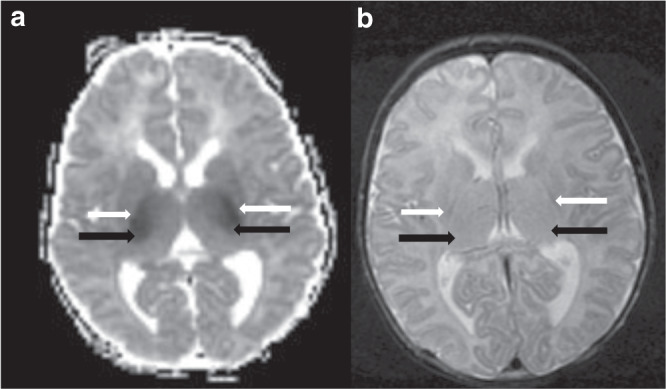
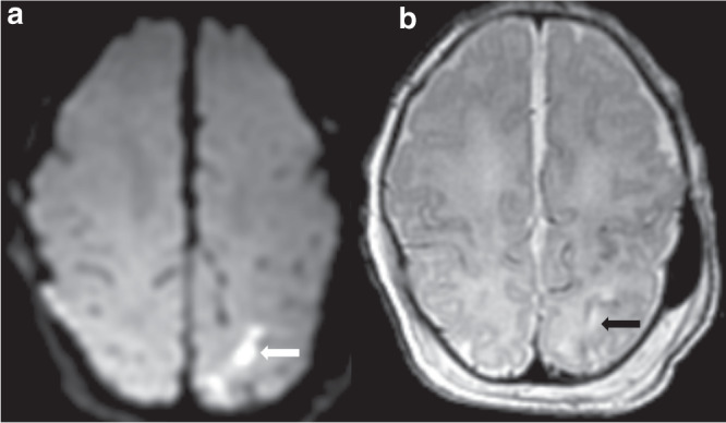
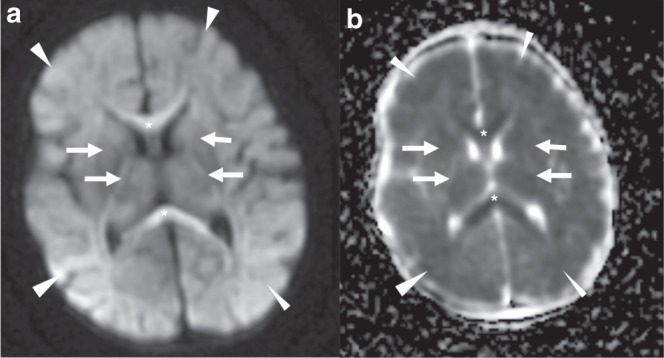
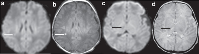
Results: Of 142 infants with mild HIE, 87 (61%) had injury on MRI at median age 5 (IQR 4-6) days. Watershed (23%), deep gray (20%) and punctate white matter (18%) injury were most common. Among the 125 (88%) infants who received a brain MRI at ≤8 days, mild (44%) injury was more common than moderate (11%) or severe (4%) injury. Subacute (37%) lesions were more commonly observed than acute (32%) or chronic lesions (1%).

Conclusion: Subacute brain injury is common in newborn infants with mild HIE. Novel neuroprotective treatments for mild HIE will ideally target both subacute and acute injury mechanisms.

Impact: Almost two-thirds of infants with mild HIE have evidence of brain injury on MRI obtained in the early neonatal period. Subacute brain injury was seen in 37% of infants with mild HIE. Neuroprotective treatments for mild HIE will ideally target both acute and subacute injury mechanisms.


Conflict of interest statement

The authors declare no competing interests.

Figures

Fig. 1
Fig. 1. Acute, deep gray pattern of hypoxic ischemic brain injury.
a ADC map demonstrating restricted diffusion involving the posterior limbs of the internal capsules (white arrows) and ventrolateral thalami (black arrows) and b T2-weighted imaging demonstrating hyperintensity involving the posterior limb of the internal capsule (white arrows) and bilateral thalami (black arrows).
Fig. 2
Fig. 2. Acute arterial watershed pattern of hypoxic ischemic brain injury.
a DWI demonstrating restricted diffusion (white arrow) and b T2 demonstrating hyperintensity (black arrow) in the left superior parietal white matter.
Fig. 3
Fig. 3. Acute, severe deep gray and watershed pattern of injury.
DWI (a) and ADC map (b) demonstrate restricted diffusion involving the deep gray (white arrows denoting reduced diffusion in the putamen, globi paladi, ventrolateral thalamus, posterior limb of the internal capsule) and watershed pattern (white arrowheads denoting reduced diffusion involving the subcortical white matter), with global injury score 84. Additional reduced diffusion involving the genu and splenium of the corpus callosum (asterisks) is compatible with pre-Wallerian degeneration from intramyelinic edema.
Fig. 4
Fig. 4. Multiple injury patterns and acuities of injury.
Acute punctate white matter injury (white arrows) with reduced diffusion (a) and T1 hyperintensity (b). Additional subacute injury to the right thalamus (black arrows) with no reduced diffusion (c) and T2 hyperintensity (d), and without volume loss.

References

    1. Kurinczuk JJ, White-Koning M, Badawi N. Epidemiology of neonatal encephalopathy and hypoxic-ischaemic encephalopathy. Early Hum. Dev. 2010;86:329–338. doi: 10.1016/j.earlhumdev.2010.05.010. - DOI - PubMed
    1. Sarnat HB, Sarnat MS. Neonatal encephalopathy following fetal distress: a clinical and electroencephalographic study. Arch. Neurol. 1976;33:696–705. doi: 10.1001/archneur.1976.00500100030012. - DOI - PubMed
    1. Robertson C, Finer N. Term infants with hypoxic-ischemic encephalopathy: outcome at 3.5 years. Dev. Med. Child Neurol. 1985;27:473–484. doi: 10.1111/j.1469-8749.1985.tb04571.x. - DOI - PubMed
    1. Azzopardi DV, et al. Moderate hypothermia to treat perinatal asphyxial encephalopathy. N. Engl. J. Med. 2009;361:1349–1358. doi: 10.1056/NEJMoa0900854. - DOI - PubMed
    1. Shankaran S, et al. Whole-body hypothermia for neonates with hypoxic-ischemic encephalopathy. N. Engl. J. Med. 2005;353:1574–1584. doi: 10.1056/NEJMcps050929. - DOI - PubMed

Publication types