

Comment
doi: 10.1038/s41422-022-00653-7.
Cuproptosis: a copper-triggered modality of mitochondrial cell death
Affiliations
- PMID: 35354936
- PMCID: PMC9061796
- DOI: 10.1038/s41422-022-00653-7
Item in Clipboard
Comment
Cuproptosis: a copper-triggered modality of mitochondrial cell death
Cell Res.
2022 May.
No abstract available
Figures
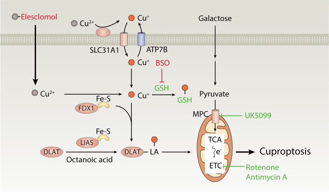
Elesclomol binds copper (Cu2+) in the extracellular environment and transports it to intracellular compartments. Increased Cu accumulation causes cuproptosis mainly through FDX1-mediated mitochondrial proteotoxic stress. On the one hand, FDX1 reduces Cu2+ to Cu+, facilitating the lipoylation (LA) and aggregation of enzymes (especially DLAT) involved in the regulation of mitochondrial TCA cycle. On the other hand, FDX1 causes the destabilization of Fe–S cluster proteins. In addition to Cu ionophores, Cu importers (e.g., SLC31A1) and exporters (e.g., ATP7B) regulate cuproptosis sensitivity by affecting intracellular Cu+ levels. GSH functions as a thiol-containing copper chelator that blocks cuproptosis, whereas BSO promotes cuproptosis by depleting GSH. The mitochondrial pyruvate carrier (MPC) inhibitor UK5099 and electron transport chain (ETC) complex I/III inhibitors (e.g., rotenone and antimycin A) attenuate elesclomol-induced cuproptosis.
Comment on
-
Copper induces cell death by targeting lipoylated TCA cycle proteins.Science. 2022 Mar 18;375(6586):1254-1261. doi: 10.1126/science.abf0529. Epub 2022 Mar 17. Science. 2022. PMID: 35298263 Free PMC article.
References
Publication types
MeSH terms
Substances
LinkOut - more resources
Full Text Sources
