Review
. 2022 Nov;22(11):701-712.
doi: 10.1038/s41577-022-00704-5. Epub 2022 Mar 30.

Heterogeneity of type 2 innate lymphoid cells

Affiliations
Review

Heterogeneity of type 2 innate lymphoid cells

Hergen Spits et al. Nat Rev Immunol. 2022 Nov.

Abstract

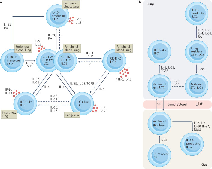
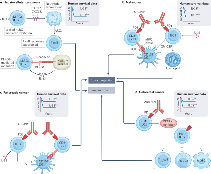
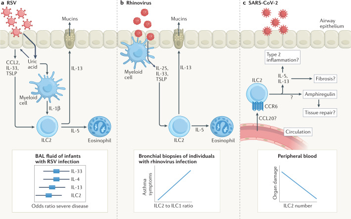
More than a decade ago, type 2 innate lymphoid cells (ILC2s) were discovered to be members of a family of innate immune cells consisting of five subsets that form a first line of defence against infections before the recruitment of adaptive immune cells. Initially, ILC2s were implicated in the early immune response to parasitic infections, but it is now clear that ILC2s are highly diverse and have crucial roles in the regulation of tissue homeostasis and repair. ILC2s can also regulate the functions of other type 2 immune cells, including T helper 2 cells, type 2 macrophages and eosinophils. Dysregulation of ILC2s contributes to type 2-mediated pathology in a wide variety of diseases, potentially making ILC2s attractive targets for therapeutic interventions. In this Review, we focus on the spectrum of ILC2 phenotypes that have been described across different tissues and disease states with an emphasis on human ILC2s. We discuss recent insights in ILC2 biology and suggest how this knowledge might be used for novel disease treatments and improved human health.


Conflict of interest statement

H.S. is a consultant for GlaxoSmithKline. J.M. declares no competing interests.

Figures

Fig. 1
Fig. 1. The plasticity and migration of ILC2s generate phenotypic diversity in tissues.
a | KLRG1+ immature type 2 innate lymphoid cells (ILC2s) in humans can generate IL-10-producing regulatory ILC2s as well as IL-5-producing and IL-13-producing ILC2s (CRTH2+CD117+ ILC2s and CRTH2+CD117 ILC2s), depending on the tissue microenvironment and cytokine milieu. It is currently unknown if IL-10-producing ILC2s can lose the capacity for IL-10 production and become IL-5-producing and IL-13-producing ILC2s. Upon activation with IL-33 and thymic stromal lymphopoietin (TSLP), human ILC2s generate CD45RO+ ILC2s that are similar to activated gut ILC2s in mice, expressing CD45RO, BATF, IRF4 and increased levels of IL-5 and IL-13. CD117 ILC2s represent committed ILC2s, but it is currently unclear if such cells are the precursors of CD45RO+ ILC2s or if CD45RO+ ILC2s can revert to CD45ROCD117 ILC2s. ILC2s, preferentially those expressing high levels of CD117, can generate ILC3-like cells producing IL-17 under the influence of IL-1β, IL-23 and transforming growth factor-β (TGFβ), or ILC1-like cells producing interferon-γ (IFNγ) under the influence of IL-1β and IL-12. Such transdifferentiation is prevented, in both cases, by IL-4. It is currently unclear if ILC2–ILC1 plasticity involves an intermediate ILC3-like stage. b | In a mouse model of Nippostrongylus brasiliensis infection, IL-25-activated ‘inflammatory’ ILC2s in the gut can migrate via the lymphatic system and blood circulation to the lung in a sphingosine 1-phosphate (S1P)-dependent manner, adding to the pool of lung-resident ST2+ ILC2s. Gut-derived ILC2s can also generate ILC3-like cells in the lung in response to type 3 cytokines such as IL-6, IL-23 and TGFβ. Gut-derived ILC2s from the lung likely return to the gut, as such cells cannot be found in the lung after parasite clearance. Additionally, lung-derived, activated ST2+ ILC2s can leave the lung and enter the circulation but it is so far unclear where such cells migrate. IL-10-producing ILC2s can be generated in both the gut and lung but the migratory behaviour of these cells is still unclear. The migratory pattern of human ILC2s is unclear but data support the recirculation of ILC2s between blood and lung in individuals with asthma. NMU, neuromedin U; RA, retinoic acid.
Fig. 2
Fig. 2. ILC2s can contribute to tumour rejection or tumour growth.
In mouse models of hepatocellular carcinoma (part a), loss of KLRG1-mediated inhibition in a population of KLRG1 type 2 innate lymphoid cells (ILC2s) results in the production of IL-13 as well as of the chemokines CXCL2 and CXCL8. CXCL2 and CXCL8 recruit neutrophils to the tumour site, which, under the influence of IL-13, produce the immunosuppressive factor arginase 1 (ARG1). ARG1 suppresses T cell responses, ultimately promoting tumour growth. In mouse models of melanoma (part b) and pancreatic cancer (part c), IL-33 induces ILC2s that subsequently activate CD103+ dendritic cells (DCs) to prime antitumour CD8+ T cells. In the case of melanoma, the production of granulocyte–macrophage colony-stimulating factor (GM-CSF) by ILC2s also recruits eosinophils to promote tumour rejection. Anti-PD1 therapy blocks cell-intrinsic inhibition of CD8+ T cells and ILC2s through the checkpoint molecule PD1 to further enhance tumour rejection. In mouse models of colorectal cancer (part d), PD1+ ILC2s enhance tumour growth, for example through the inhibition of natural killer (NK) cell responses and enhancing the responses of regulatory T (Treg) cells and myeloid-derived suppressor cells (MDSCs). Inhibition of PD1 signalling in ILC2s, through either pharmacological or genetic inhibition of the peroxisome proliferator-activated receptor-γ (PPARγ) pathway (which reduces PD1 expression) or anti-PD1 therapy promotes tumour rejection in colorectal cancer. These mechanistic insights from mouse models are supported by data derived from human cancer tissues (see inset schematic graphs). Genome-wide transcriptional data indicative of high intratumoural levels of IL33 (in hepatocellular carcinoma (part a) and pancreatic cancer (part c)) or high levels of ILC2-associated gene expression (in melanoma (part b) and colorectal cancer (part d)) predict better survival in humans.
Fig. 3
Fig. 3. ILC2s are involved in adipose tissue homeostasis.
In mouse adipose tissue, sympathetic nerves and alternatively activated, M2-type macrophages produce noradrenaline that binds β-adrenergic receptors on mesenchymal adipose progenitor cells. This leads to their production of IL-33 and glial cell-derived neurotrophic factor (GDNF), for which the receptors ST2 and RET, respectively, are expressed on adipose tissue type 2 innate lymphoid cells (ILC2s). Activation of adipose tissue ILC2s leads to IL-4 and IL-13 production, which act on mesenchymal adipose cells to promote their production of the eosinophil chemotactic factor CCL11 (also known as eotaxin). Together with ILC2-derived IL-5, this recruits eosinophils to adipose tissue and supports their maintenance. Eosinophils produce IL-4 that supports the differentiation of alternatively activated macrophages, thereby creating a positive-feedback loop for a type 2 environment in the adipose tissue. Adipose tissue ILC2 activation also leads to the production of Met-Enkephalin (MetEnk), which acts on opioid receptors on adipocytes, resulting in a process known as beiging that reduces insulin resistance, a component of the metabolic syndrome.
Fig. 4
Fig. 4. ILC2s are involved in virus-induced exacerbations of airway inflammation.
a | In a mouse model, respiratory syncytial virus (RSV) infection induces activation of type 2 innate lymphoid cells (ILC2s) in the airways via uric acid-mediated production of IL-33, thymic stromal lymphopoietin (TSLP) and CCL2 by airway epithelial cells, as well as IL-1β production by myeloid cells. IL-13 production by ILC2s mediates mucin production as well as other downstream effects associated with asthma such as the IL-5-mediated maintenance of eosinophils. High levels of IL-33, IL-4, IL-13 and ILC2s in bronchoalveolar lavage (BAL) fluid are associated with more severe airway inflammation in infants with RSV infection. b | In rhinovirus infection in mice, alarmins such as IL-33, produced by airway epithelial cells and myeloid cells, drive ILC2 activation, leading to IL-5 and IL-13 production for the recruitment of eosinophils and mucin production, respectively. In experimental rhinovirus infection in humans, the bronchial ILC2 to ILC1 ratio correlates with asthma symptom score. c | In severe acute respiratory syndrome coronavirus 2 (SARS-CoV-2) infection in humans, numbers of CCR6+ ILC2s in the circulation are reduced, which correlates with serum levels of CCL20, indicative of the CCL20–CCR6-dependent recruitment of ILC2s from the circulation to sites of inflammation such as the lungs. Blood ILC2 numbers are inversely correlated with serum markers of organ damage in patients with coronavirus disease 2019 (COVID-19), suggesting a potential detrimental role of ILC2s in inflamed tissues. However, the precise role for ILC2s in the airways, including their production of cytokines, such as amphiregulin, which might be involved in lung tissue repair, in patients with COVID-19 remains unclear.

References

    1. Neill DR, et al. Nuocytes represent a new innate effector leukocyte that mediates type-2 immunity. Nature. 2010;464:1367–1370. doi: 10.1038/nature08900. - DOI - PMC - PubMed
    1. Moro K, et al. Innate production of TH2 cytokines by adipose tissue-associated c-Kit+Sca-1+ lymphoid cells. Nature. 2010;463:540–544. doi: 10.1038/nature08636. - DOI - PubMed
    1. Price AE, et al. Systemically dispersed innate IL-13-expressing cells in type 2 immunity. Proc. Natl Acad. Sci. USA. 2010;107:11489–11494. doi: 10.1073/pnas.1003988107. - DOI - PMC - PubMed
    1. Mjosberg JM, et al. Human IL-25- and IL-33-responsive type 2 innate lymphoid cells are defined by expression of CRTH2 and CD161. Nat. Immunol. 2011;12:1055–1062. doi: 10.1038/ni.2104. - DOI - PubMed
    1. Monticelli LA, et al. Innate lymphoid cells promote lung-tissue homeostasis after infection with influenza virus. Nat. Immunol. 2011;12:1045–1054. doi: 10.1038/ni.2131. - DOI - PMC - PubMed