. 2022 Mar 9:9:808036.
doi: 10.3389/fmolb.2022.808036. eCollection 2022.

Human Mitochondrial DNA Polymerase Metal Dependent UV Lesion Bypassing Ability

Affiliations

Human Mitochondrial DNA Polymerase Metal Dependent UV Lesion Bypassing Ability

Joon Park et al. Front Mol Biosci. .

Abstract

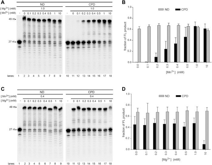
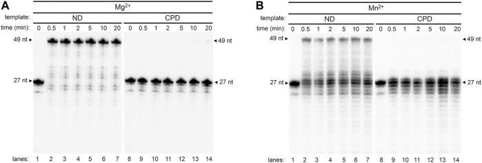
Human mitochondrial DNA contains more UV-induced lesions than the nuclear DNA due to lack of mechanism to remove bulky photoproducts. Human DNA polymerase gamma (Pol γ) is the sole DNA replicase in mitochondria, which contains a polymerase (pol) and an exonuclease (exo) active site. Previous studies showed that Pol γ only displays UV lesion bypassing when its exonuclease activity is obliterated. To investigate the reaction environment on Pol γ translesion activity, we tested Pol γ DNA activity in the presence of different metal ions. While Pol γ is unable to replicate through UV lesions on DNA templates in the presence of Mg2+, it exhibits robust translesion DNA synthesis (TLS) on cyclobutane pyrimidine dimer (CPD)-containing template when Mg2+ was mixed with or completely replaced by Mn2+. Under these conditions, the efficiency of Pol γ's TLS opposite CPD is near to that on a non-damaged template and is 800-fold higher than that of exonuclease-deficient Pol γ. Interestingly, Pol γ exhibits higher exonuclease activity in the presence of Mn2+ than with Mg2+, suggesting Mn2+-stimulated Pol γ TLS is not via suppressing its exonuclease activity. We suggest that Mn2+ ion expands Pol γ's pol active site relative to Mg2+ so that a UV lesion can be accommodated and blocks the communication between pol and exo active sites to execute translesion DNA synthesis.

Keywords: DNA polymerase gamma; TLS; UV lesion; metal-dependence; mitochondrial DNA.


Conflict of interest statement

The authors declare that the research was conducted in the absence of any commercial or financial relationships that could be construed as a potential conflict of interest.

Figures

FIGURE 1
FIGURE 1
DNA synthesis of wild-type Pol γ on non-damaged and CPD-containing templates in the presence of Mg2+ or Mn2+. (A) Structure of thymine bases, CPD, and (6-4)PP where nitrogen is represented in blue spheres, oxygen in red spheres, carbon in black spheres, and phosphorus in golden spheres. (B) Time-dependent DNA synthesis of wild-type Pol γ on 5′-32P-Primer N (PN) annealed to a non-damaged template (T-ND) (lanes 1–8) or CPD-containing template (T-CPD) (lanes 9–16) in the presence of Mg2+. (C) Time-dependent DNA synthesis of wild-type Pol γ on 5′-32P-PN annealed to T-ND (lanes 1–8) or T-CPD (lanes 9–16) in the presence of Mn2+. (D) Quantification of normalized full-length (FL) products from (B) and (C) over time in seconds. The results are presented as mean and standard deviation from three independent experiments.
FIGURE 2
FIGURE 2
DNA synthesis of exonuclease-deficient Pol γ on non-damaged and CPD-containing templates in the presence of Mg2+ or Mn2+. (A) Time-dependent DNA synthesis of exonuclease-deficient (exo-) Pol γ on 5′-32P-PN annealed to T-ND in the presence of Mg2+ (lanes 1–8) or Mn2+ (lanes 9–16) (B) Time-dependent DNA synthesis of Pol γ exo-on 5′-32P-PN annealed to T-CPD in the presence of Mg2+ (lanes 1–8) or Mn2+ (lanes 9–16). (C) Quantification of normalized full-length (FL) products from (A) and (B) over time in seconds. The results are presented as mean and standard deviation from three independent experiments.
FIGURE 3
FIGURE 3
Metal-dependent DNA synthesis of wild-type and exonuclease-deficient Pol γ on a (6-4)PP-containing template. Time-dependent polymerization assay of wild-type Pol γ (exo+) on 5′-32P-PN annealed to T-(6–4)PP in the presence of Mg2+ (lanes 2 and 3) or in the presence of Mn2+ (lanes 4 and 5) and of Pol γ (exo-) on identical DNA substrates in the presence of Mg2+ (lanes 7 and 8) or Mn2+ (lanes 9 and 10).
FIGURE 4
FIGURE 4
Metal-regulated exonuclease activity of wild-type Pol γ. (A) Exonuclease assay of Pol γ on a single-stranded DNA, 5′-32P-PN, in the presence of Mg2+ (lanes 1–8) or in the presence of Mn2+ (lanes 9–16). (B) Exonuclease assay Pol γ on primer/template, 5′-32P-PN/T-ND, in the presence of Mg2+ (lanes 1–8) or in the presence of Mn2+ (lanes 9–16) (C). Quantification of normalized fraction of substrate remaining in (A) and (B) over time in seconds shown. The results are presented as mean and standard deviation from three independent experiments. Shorter time points (0–200 s) are shown as an inset.
FIGURE 5
FIGURE 5
Effect of a single metal ion on wild-type Pol γ DNA activity on non-damaged and CPD-containing templates. (A) Wild-type Pol γ primer extension on T-ND annealed to 5′-32P-PN in the presence of Mg2+(lanes 1–9) or Mn2+ (lanes 10-18). (B) Quantification of the full-length product on T-ND vs. a gradient of Mg2+ or Mn2+ shown in (A). (C) Wild-type Pol γ primer extension on T-CPD annealed to 5′-32P-PN in the presence of Mg2+ (lanes 1–9) or Mn2+ (lanes 10-18). (D) Quantification of the full-length product on T-CPD vs. a gradient of Mg2+ or Mn2+ shown in (C).
FIGURE 6
FIGURE 6
Effect of mixed metal ions on wild-type Pol γ DNA synthesis on non-damaged and CPD-containing templates in a mixture of Mg2+ and Mn2+. (A) Wild-type Pol γ primer extension on T-ND (lanes 1–9) or T-CPD (lanes 10–18) annealed to the 5′-32P-PN at a constant concentration of Mg2+ and varied concentrations of Mn2+. (B) Quantification of the full-length product vs. concentration of Mn2+ shown in (A). (C) Wild-type Pol γ primer extension on T-ND (lanes 1–9) or T-CPD (lanes 10–18) annealed to the 5′-32P-PN at constant concentration of Mn2+ and varied concentrations of Mg2+. (D) Quantification of the full-length product vs. concentration of Mg2+ shown in (C).
FIGURE 7
FIGURE 7
Evaluation of wild-type T7 polymerase activity on a non-damaged and CPD substrates in the presence of Mg2+ or Mn2+. (A) Time course activity assay of wild-type T7 DNA polymerase on T-ND (lanes 1–8) and on T-CPD (lanes 9–16) annealed to 5′- 32P-PN in the presence of Mg2+. (B) Time course activity assay of wild-type T7 DNA polymerase on T-ND (lanes 1–8) and on T-CPD (lanes 9–16) annealed to 5′-32P-PN in the presence of Mn2+.
FIGURE 8
FIGURE 8
Comparison of pol site conformation of Pol η and Pol γ. Left, Pol η ternary complex with CPD (red)-containing template (blue) and primer (magenta) and incoming nucleotide (green) (PDB: 3MR3). Middle, modeled Pol γ-CPD containing primer/template DNA showing the CPD (red) steric clashes with the O helices. Right, Pol γ ternary complex with primer/template and incoming nucleotide showing the bending of the template by O- and O1-helix.
FIGURE 9
FIGURE 9
Schematic model of the hypothetical function of Mg2+ and Mn2+ in reshaping Pol γ′s active site. Illustration of Pol γ (green) on an ND- (blue trapezoid) or CPD-containing template (violet rhomboid). (A) Representation of DNA replication by Pol γ on an ND-containing substrate in the presence of Mg2+, Mg2+/Mn2+, or Mn2+ showing full extension in all cases without significant changes in the catalytic site (yellow trapezoid). (B) Representation of DNA replication by Pol γ on a CPD-containing template illustrating a change of the Pol γ′s catalytic site (yellow trapezoid) in the presence of Mn2+ upon encountering CPD to trigger TLS activity.

Similar articles

Cited by

References

    1. Bębenek A., Ziuzia-Graczyk I. (2018). Fidelity of DNA Replication-A Matter of Proofreading. Curr. Genet. 64, 985–996. 10.1007/s00294-018-0820-1 - DOI - PMC - PubMed
    1. Beckman R. A., Mildvan A. S., Loeb L. A. (1985). On the Fidelity of DNA Replication: Manganese Mutagenesis In Vitro . Biochemistry 24, 5810–5817. 10.1021/bi00342a019 - DOI - PubMed
    1. Bess A. S., Crocker T. L., Ryde I. T., Meyer J. N. (2012). Mitochondrial Dynamics and Autophagy Aid in Removal of Persistent Mitochondrial DNA Damage in Caenorhabditis elegans . Nucleic Acids Res. 40, 7916–7931. 10.1093/nar/gks532 - DOI - PMC - PubMed
    1. Bess A. S., Ryde I. T., Hinton D. E., Meyer J. N. (2013). UVC-induced Mitochondrial Degradation via Autophagy Correlates with mtDNA Damage Removal in Primary Human Fibroblasts. J. Biochem. Mol. Toxicol. 27, 28–41. 10.1002/jbt.21440 - DOI - PMC - PubMed
    1. Bharti S. K., Sommers J. A., Zhou J., Kaplan D. L., Spelbrink J. N., Mergny J.-L., et al. (2014). DNA Sequences Proximal to Human Mitochondrial DNA Deletion Breakpoints Prevalent in Human Disease Form G-Quadruplexes, a Class of DNA Structures Inefficiently Unwound by the Mitochondrial Replicative Twinkle Helicase. J. Biol. Chem. 289, 29975–29993. 10.1074/jbc.m114.567073 - DOI - PMC - PubMed

LinkOut - more resources