

Acute disseminated encephalomyelitis (ADEM) after SARS- CoV-2 vaccination: A case report
- PMID: 35355527
- PMCID: PMC8957291
- DOI: 10.1016/j.radcr.2022.03.013
Acute disseminated encephalomyelitis (ADEM) after SARS- CoV-2 vaccination: A case report
Abstract
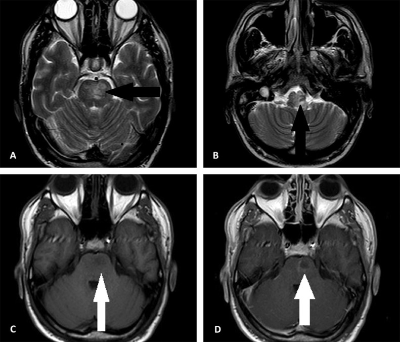
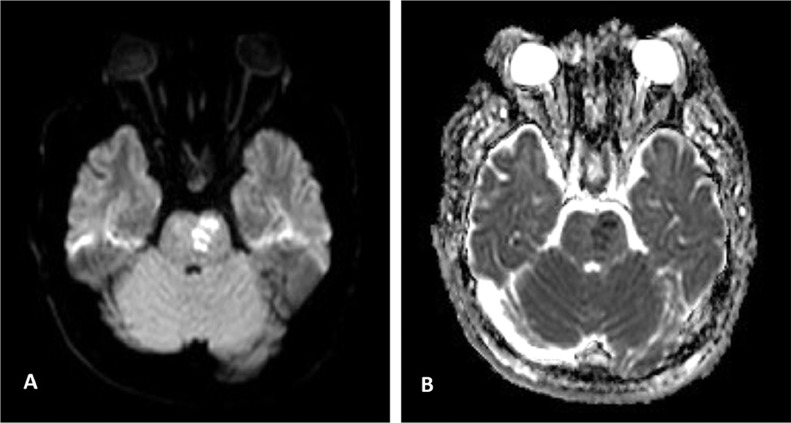
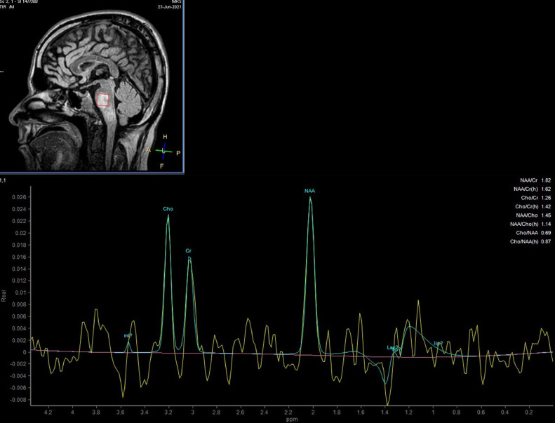
Acute disseminated encephalomyelitis (ADEM) is an acute demyelinating disorder of the central nervous system that is ordinarily monophasic. ADEM can develop following infection or vaccination. Here, we present a 37 y/o male patient with progressive muscle weakness in all limbs along with dysphagia following COVID-19 vaccination. Brain magnetic resonance imaging (MRI) revealed typical imaging findings which presented as multifocal T2-FLAIR signal changes in the corticospinal tract, pons, and temporal lobe with diffusion restriction. Magnetic resonance spectroscopy (MRS) further confirmed the diagnosis by the typical elevation of the Choline and Myoinositol peaks. Neurologic impairments have been reported as the potential side effects of COVID-19 vaccines. Appropriate imaging modalities together with a thorough clinical examination are essential for making a correct diagnosis.
Keywords: ADEM; Brain ,MRI ,MRS; Demyelinating disease.
© 2022 Published by Elsevier Inc. on behalf of University of Washington.
Figures
References
-
- Alper G. Acute disseminated encephalomyelitis. J Child Neurol. 2012;27(11):1408–1425. - PubMed
-
- Pohl D., Alper G., Van Haren K., Kornberg A.J., Lucchinetti C.F., Tenembaum S., et al. Acute disseminated encephalomyelitis: updates on an inflammatory CNS syndrome. Neurology. 2016;87(9 Supplement 2):S38–S45. - PubMed
-
- Tenembaum S., Chitnis T., Ness J., Hahn J.S. Group ftIPMS. Acute disseminated encephalomyelitis. Neurology. 2007;68(16 suppl 2):S23–S36. - PubMed
-
- Cole J., Evans E., Mwangi M., Mar S. Acute disseminated encephalomyelitis in children: an updated review based on current diagnostic criteria. Pediatr Neurol. 2019;100:26–34. - PubMed
Publication types
LinkOut - more resources
Full Text Sources
Miscellaneous
