

Spontaneous round ligament hematoma as an unusual cause of pelvic pain in a young female patient: MRI demonstration
- PMID: 35355531
- PMCID: PMC8958461
- DOI: 10.1016/j.radcr.2022.02.079
Spontaneous round ligament hematoma as an unusual cause of pelvic pain in a young female patient: MRI demonstration
Erratum in
-
Corrigendum to "Spontaneous round ligament hematoma as an unusual cause of pelvic pain in a young female patient: MRI demonstration" [Radiology Case Reports 17 (2022) 1765-1769].Radiol Case Rep. 2022 Jul 8;17(9):3446. doi: 10.1016/j.radcr.2022.06.023. eCollection 2022 Sep. Radiol Case Rep. 2022. PMID: 35909931 Free PMC article.
Abstract
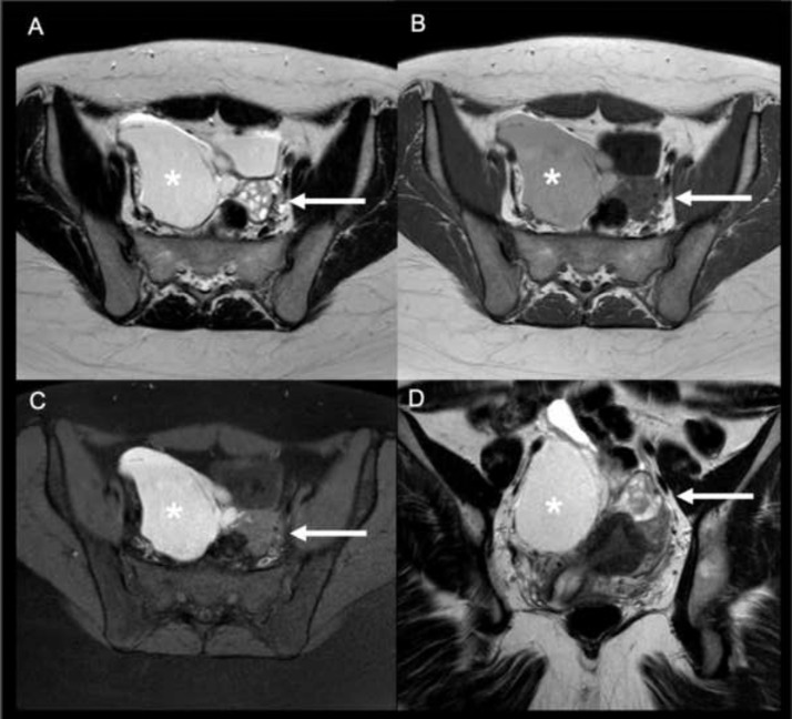
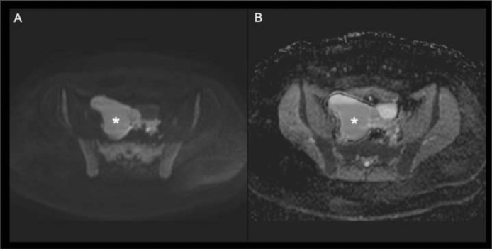
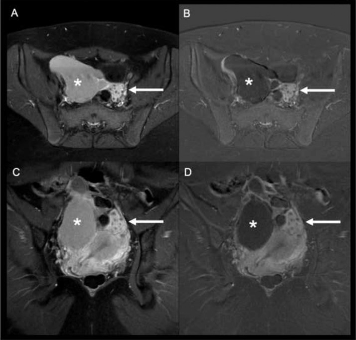
The cause of pelvic pain remains a significant diagnostic challenge, even for experienced radiologists. An accurate differential diagnosis has to be done according to the patient's age and gender. Spontaneous round ligament hematoma is an uncommon cause of acute pelvic pain in adult female patients. To the best of our knowledge, it has never been reported in the literature in the paediatric population. Ultrasound examination is the first line imaging modality for pelvic pain evaluation in young women but it might result inconclusive. Thanks to its panoramic view and multiparametric approach, the MRI can play a pivotal role in the diagnosis of spontaneous round ligament hematoma in paediatric female patients, resulting in a more effective patient's therapeutic management.
Keywords: MRI; Pediatric pelvic pain; Spontaneous round ligament hematoma.
© 2022 The Authors. Published by Elsevier Inc. on behalf of University of Washington.
Figures
References
-
- Borrero E, Weil P H, Cooper J J. Hemorrhage into the round ligament. JAMA. 1983 Jun 24;249(24):3306. - PubMed
Publication types
LinkOut - more resources
Full Text Sources
