. 2022 Mar 9:13:851401.
doi: 10.3389/fphar.2022.851401. eCollection 2022.

HIF1α/VEGF Feedback Loop Contributes to 5-Fluorouracil Resistance

Affiliations

HIF1α/VEGF Feedback Loop Contributes to 5-Fluorouracil Resistance

Bin Shi et al. Front Pharmacol. .

Abstract

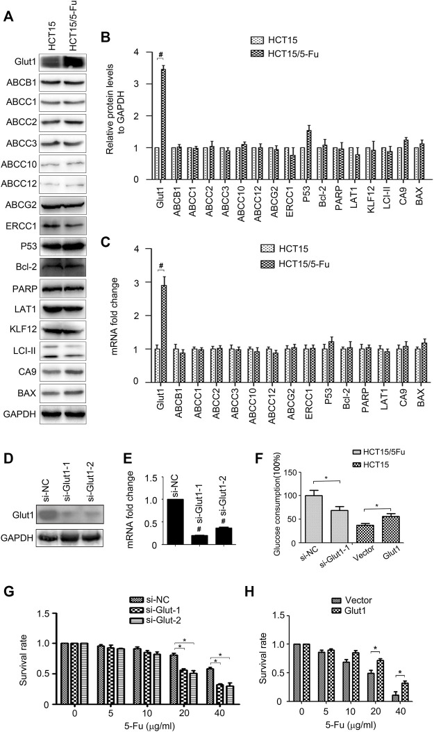
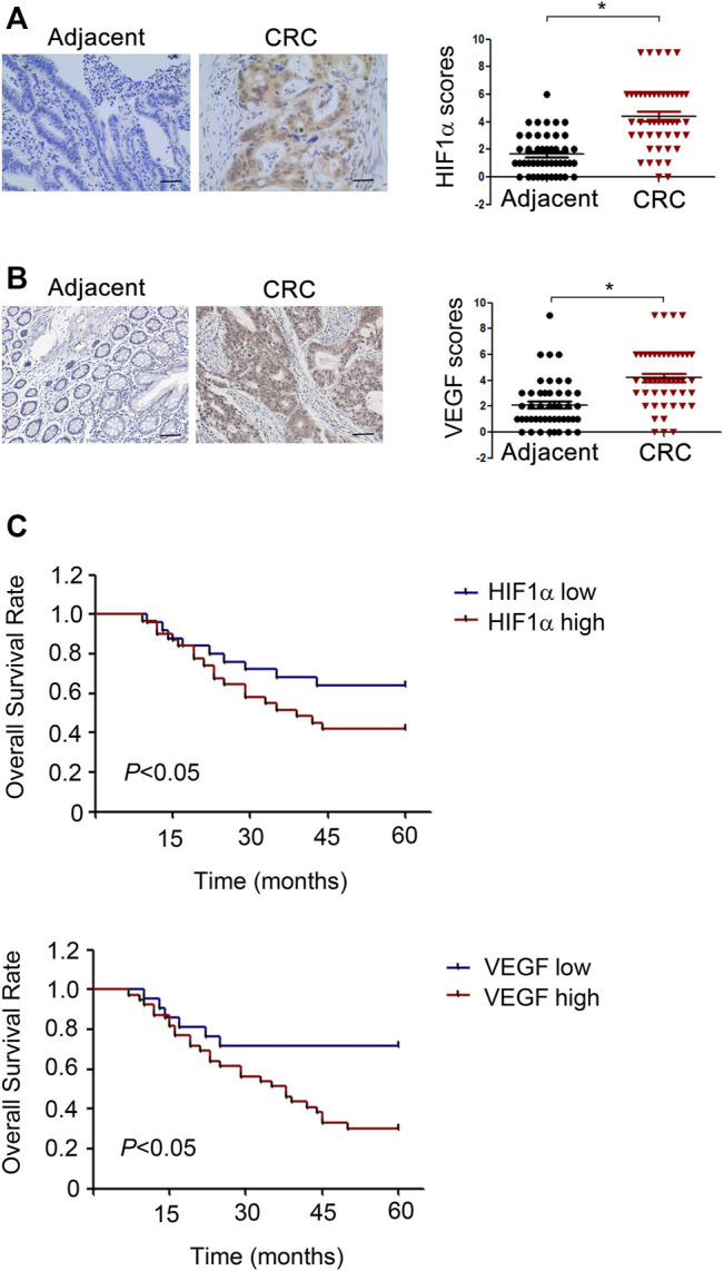
5-Fluorouracil (5-Fu) is one of the basic drugs in colorectal cancer (CRC) chemotherapy, and its efficacy is mainly limited by the acquisition of drug resistance. However, the underlying mechanisms remain unclear. In this study, hypoxia inducible factor 1α (HIF1α) was screened for high expression in 5-Fu resistant HCT115 cells, which displayed epithelial-mesenchymal transition (EMT) phenotype. Suppression of HIF1α reversed EMT phenotype, reduced glucose transporter 1 (Glut1) expression, a key molecule mediated drug resistance. Moreover, we unveiled that vascular endothelial growth factor (VEGF) was regulated by HIF1α and mediated HIF1α-maintained malignant phenotype of 5-Fu resistant cells. Further studies verified that AKT/GSK3β signaling was activated in resistant cells and controlled HIF1α expression. Interestingly, we demonstrated that VEGF could feedback up-regulate HIF1α via AKT/GSK3β signaling. Clinically, HIF1α and VEGF were high expressed and associated with survival and prognosis in CRC patients. In conclusion, our findings proposed that HIF1α/VEGF feedback loop contributed to 5-Fu resistance, which might be potential therapeutic targets.

Keywords: Akt/GSK3β; EMT; GLUT1; HIF1α; VEGF; drug resistance.


Conflict of interest statement

The authors declare that the research was conducted in the absence of any commercial or financial relationships that could be construed as a potential conflict of interest.

Figures

FIGURE 1
FIGURE 1
5-Fu resistant CRC cells acquire EMT phenotype. (A) HCT15 and HCT15/5-Fu cells were treated with increasing concentrations of 5-Fu for 48 h. CCK8 assay was used to quantify the viable cells. (B) Morphology of HCT15 and HCT15/5-Fu cells. (C) Transwell assay was performed to measure the migration of HCT15 and HCT15/5-Fu cells. (D,E) The protein and mRNA expression of E-cadherin and Vimentin in HCT15 and HCT15/5-Fu cells were examined by WB and RT-PCR, respectively. (F) The expression and cellular localization of E-cadherin and Vimentin were detected by IF staining. Nuclei were visualized with DAPI staining. Data represented as mean ± SD were from three independent experiments. *: p < 0.05, #: p < 0.01.
FIGURE 2
FIGURE 2
Glut1 maintains the resistance phenotype of 5-Fu resistant CRC cells. (A,B) The protein expression of Glut1, ABCB1, ABCC1, ABCC2, ABCC3, ABCC10, ABCC12, ABCG2, ERCC1, P53, Bcl-2, PARP, LAT1, KLF12, LCI-II, CA9 and BAX in HCT15 and HCT15/5-Fu cells were examined by WB. GAPDH servers as the loading control. (C) The mRNA expression of Glut1, ABCB1, ABCC1, ABCC2, ABCC3, ABCC10, ABCC12, ABCG2, ERCC1, P53, Bcl-2, PARP, LAT1, CA9 and BAX in in HCT15 and HCT15/5-Fu cells were examined by RT-PCR. (D,E) Expression of Glut1 protein and mRNA in HCT15/5-Fu cells transfected with si-NC or si-Glut1 for 24 h were detected by WB and quantitative RT-PCR, respectively. (F) Glucose concentration in the supernatants was measured in HCT15/5-Fu cells transfected with si-NC or si-Glut1 and HCT15 cells transfected with control vector plasmid or pcDNA-Glut1. (G) HCT15/5-Fu cells were transfected with si-NC or si-Glut1 for 24 h were treated with increasing concentrations of 5-Fu for 48 h. CCK8 assay was used to quantify the viable cells. (H) HCT15 cells were transfected with control vector plasmid or pcDNA3.1-Glut1 for 24 h were treated with increasing concentrations of 5-Fu for 48 h. CCK8 assay was used to quantify the viable cells. Data represented as mean ± SD were from three independent experiments. *: p < 0.05, #: p < 0.01. si-NC: negative control siRNA.
FIGURE 3
FIGURE 3
HIF1α is up-regulated in 5-Fu resistant CRC cells. (A–C) The protein and mRNA expression of Snail, Slug, Twist, ZEB1, β-catenin and HIF1α in HCT15 and HCT15/5-Fu cells were examined by WB and RT-PCR, respectively. (D) The HIF1α expression in cytoplasm and nucleus in HCT15 and HCT15/5-Fu cells were examined by WB. (E) The cellar localization of HIF1α in HCT15 and HCT15/5-Fu cells was analyzed by IF staining. Nuclei were visualized with DAPI staining. #: p < 0.01.
FIGURE 4
FIGURE 4
HIF1α regulates VEGF expression in CRC cells. (A) the mRNA expression of TGF-β, TNF-α, VEGF, EGF, PDGF, b-FGF, HGF, and IGF in HCT15 cells transfected with si-NC or si-HIF1α for 48 h were detected by RT-PCR. (B) the protein expression of VEGF in HCT15 cells transfected with si-NC or si-HIF1α for 48 h were detected by WB. (C,D) the mRNA and protein expression of VEGF in HCT15 and HCT15/5-Fu cells were detected by WB and RT-PCR, respectively. (E,F) the mRNA and protein expression of VEGF in HCT15/5-Fu cells transfected with si-NC or si-HIF1α for 48 h were detected by WB and RT-PCR, respectively. #: p < 0.01, *: p < 0.05.
FIGURE 5
FIGURE 5
VEGF mediates HIF1α-maintained the aggressive phenotype of 5-Fu resistant CRC cells. (A,B) Expression of HIF1α, E-cadherin, Vimentin, and Glut1 protein and mRNA in HCT15/5-Fu cells transfected with si-NC, si-HIF1α+vector or si-HIF1α+VEGF for 48 h were detected byWB and RT-PCR, respectively. (C) HCT15/5-Fu cells transfected with si-NC, si-HIF1α+control vector or si-HIF1α+VEGF overexpression plasmid for 24 h were treated with increasing concentrations of 5-Fu for 48 h. CCK8 assay was used to quantify the viable cells. (D) HCT15/5-Fu cells were transfected with si-NC, si-HIF1α+vector or si-HIF1α+VEGF for 48 h, the migration capability was detected by transwell assay. Data represented as mean ± SD were from three independent experiments. *: p < 0.05.
FIGURE 6
FIGURE 6
AKT/GSK3β signal is crucial for the aggressive phenotype of 5-Fu resistant CRC cells. (A) The expression of p-AKT, AKT, p-GSK3β, GSK3β, p-p65, p65, p-p38-MAPK and p38-MAPK in HCT15 and HCT15/5-Fu cells were detected by WB. (B) HCT15/5-Fu cells were treated with LY294002 (20 μM) for 24 h, the expression of p-AKT, AKT, p-GSK3β, GSK3β and HIF1α were examined by WB. (C) HCT15/5-Fu cells were treated with LY294002 (20 μM) or DMSO for 24 h, then treated with increasing concentrations of 5-Fu for 48 h. CCK8 assay was used to quantify the viable cells. (D) Transwell assay was performed to examine the migration of HCT15/5-Fu cells treated with LY294002 (20 μM) or DMSO for 24 h. Data represented as mean ± SD were from three independent experiments. *: p < 0.05, #: p < 0.01.
FIGURE 7
FIGURE 7
VEGF feedback regulates HIF1α expression in CRC cells. (A,B) The expression of p-AKT, AKT, p-GSK3β, GSK3β, p-VEGFR2, VEGFR2 and HIF1α in HCT15 and HCT116 cells treated with PBS or VEGF (10 ng/ml) for 24 h were detected by WB. *: p < 0.05, #: p < 0.01.
FIGURE 8
FIGURE 8
HIF1α and VEGF are high expressed in CRC patient tissues and predict poor prognosis. (A,B) Left, Representative IHC staining of HIF1α in CRC tumor sample and normal tissue sample. Right, Scores for HIF1α and VEGF staining in 60 pairs tumor tissue and adjacent normal tissue samples from CRC patients. (C) Univariate survival analysis of overall survival in CRC patients as determined by Kaplan-Meier plots estimates based on HIF1α and VEGF protein expression.
FIGURE 9
FIGURE 9
The graphic illustration of HIF1α/VEGF feedback loop contributes to 5-Fu resistance and metastasis in CRC.

Similar articles

Cited by

References

    1. Abad E., Graifer D., Lyakhovich A. (2020). DNA Damage Response and Resistance of Cancer Stem Cells. Cancer Lett. 474, 106–117. 10.1016/j.canlet.2020.01.008 - DOI - PubMed
    1. Apte R. S., Chen D. S., Ferrara N. (2019). VEGF in Signaling and Disease: Beyond Discovery and Development. Cell 176 (6), 1248–1264. 10.1016/j.cell.2019.01.021 - DOI - PMC - PubMed
    1. Blondy S., David V., Verdier M., Mathonnet M., Perraud A., Christou N. (2020). 5-Fluorouracil Resistance Mechanisms in Colorectal Cancer: From Classical Pathways to Promising Processes. Cancer Sci. 111 (9), 3142–3154. 10.1111/cas.14532 - DOI - PMC - PubMed
    1. Candido J., Hagemann T. (2013). Cancer-Related Inflammation. J. Clin. Immunol. 33 (Suppl. 1), S79–S84. 10.1007/s10875-012-9847-0 - DOI - PubMed
    1. Chae Y. C., Kim J. H. (2018). Cancer Stem Cell Metabolism: Target for Cancer Therapy. BMB Rep. 51 (7), 319–326. 10.5483/bmbrep.2018.51.7.112 - DOI - PMC - PubMed