. 2022 Mar 20:2022:2453617.
doi: 10.1155/2022/2453617. eCollection 2022.

Fluorofenidone Inhibits UUO/IRI-Induced Renal Fibrosis by Reducing Mitochondrial Damage

Affiliations

Fluorofenidone Inhibits UUO/IRI-Induced Renal Fibrosis by Reducing Mitochondrial Damage

Xiaohua Liao et al. Oxid Med Cell Longev. .

Abstract

Objective: Mitochondrial damage contributes to extracellular matrix (ECM) deposition and renal fibrosis. In this study, we aimed (1) to investigate whether fluorofenidone (AKF-PD) can attenuate mitochondrial damage in two renal fibrosis models: unilateral ureteral obstruction (UUO) and renal ischemia-reperfusion injury (IRI), and (2) to explore the underlying mechanism.

Method: Mitochondrial damage and renal lesions were analyzed in the UUO and IRI models. Mitochondrial energy metabolism, mitochondrial biogenesis, and oxidative stress were measured to assess the effect of AKF-PD on mitochondrial damage and to explore the underlying mechanism. In addition, HK-2 cells were stimulated with TGF-β with and without AKF-PD. The mitochondrial morphology, mtROS, ATP contents, and redox-related proteins were then examined.

Results: In both UUO and IRI models, AKF-PD relieved renal fibrosis, maintained mitochondrial structure, and increased mitochondrial DNA copy numbers. The protection was associated with (1) sustaining mitochondrial energy metabolism, evident by elevations of tricarboxylic acid (TCA) cycle enzymes and mitochondrial respiratory chain complexes; (2) improving mitochondrial biogenesis with increases of TFAM, NRF1, PGC-1α, and SIRT1; and (3) reducing mitochondrial oxidative stress likely via regulating SOD2, SIRT3, and NOX4 expressions. In HK-2 cells treated with TGF-β, AKF-PD protected mitochondria along with improving mitochondrial morphology, enhancing ATP production, reducing mtROS, and regulating SOD2, SIRT3, and NOX4 expression.

Conclusion: We demonstrate that AKF-PD inhibited renal fibrosis at least in part via protecting mitochondria from damages developed in the UUO and IRI models. The mitochondrial protection was associated with sustaining mitochondrial energy metabolism, improving mitochondrial biogenesis, and reducing mitochondrial oxidative stress. This research verified the protective effect of AKF-PD on mitochondria in the UUO and IRI models and elaborated the underlying mechanism.


Conflict of interest statement

The authors declare no conflicts of interest.

Figures

Figure 1
Figure 1
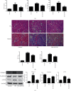
AKF-PD reduced renal fibrosis in the UUO model. (a) HE and Masson staining of renal tissues from the indicated mouse groups (n = 5); typical images (×200) for indicated groups are shown (upper panel). Quantifications for individual treatments (n = 5) are also provided (bottom panel). (b) Immunohistochemistry analyses of collagen I and collagen III in the indicated renal tissues. Typical images (×200) for individual groups and their quantifications (n = 4) are included. (c) Real-time PCR analyses for collagen I and collagen III expressions in the indicated mouse groups (n = 4). (d) The expression of FN and α-SMA in renal tissue from mice with the indicated treatments was measured by western blot. Typical images and quantification (n = 4) are shown. P < 0.05 and ∗∗P < 0.01 UUO group vs. sham group; #P < 0.05 and ##P < 0.01 UUO+AKF-PD group vs. UUO group. Data were analyzed by one-way ANOVA. AKF-PD: fluorofenidone; COL-I: collagen I; COL-III: collagen III; FN: fibronectin; α-SMA: alpha smooth muscle actin.
Figure 2
Figure 2
AKF-PD reduced renal fibrosis in the IRI model. (a) Serum urea nitrogen, creatinine, and uric acid from the indicated animal groups were determined and quantified (n = 5). (b) HE and Masson staining of renal tissues obtained from mice with the indicated treatments. Typical images (×200) and quantifications (n = 5) are shown. (c) The expression of collagen I, E-cadherin, and α-SMA in renal tissue from IRI mice was measured by western blot with typical images and quantifications (n = 4) included. P < 0.05 and ∗∗P < 0.01 IRI group vs. control group; #P < 0.05 and ##P < 0.01 IRI+AKF-PD group vs. IRI group. Data were analyzed by one-way ANOVA. AKF-PD: fluorofenidone; BUN: blood urea nitrogen; Cr: creatinine; UA: uric acid; α-SMA: alpha smooth muscle actin.
Figure 3
Figure 3
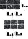
AKF-PD reduced mitochondrial damage in the UUO and IRI models. (a) Mitochondrial morphology of renal tubular epithelial cells from the indicated mouse group was examined by transmission electron microscopy. (b) ATP levels of renal tissue from the indicated mice were measured by the ATP assay kit and quantified (n = 4). (c) The expression of ND1 and ND4 in renal tissue from the indicated mice was measured by real-time PCR (n = 4). (d) Mitochondrial morphology of renal tubular epithelial cells from the indicated mouse group was examined by transmission electron microscopy. (e) The expression of ND1 and ND4 in renal tissue from the indicated mice was measured by real-time PCR (n = 4). P < 0.05 and ∗∗P < 0.01 UUO/IRI group vs. sham/control group; #P < 0.05 and ##P < 0.01 UUO+AKF-PD/IRI+AKF-PD group vs. UUO/IRI group. Data were analyzed by one-way ANOVA. AKF-PD: fluorofenidone; ND1: NADPH dehydrogenase subunit 1; ND4: NADPH dehydrogenase subunit 4.
Figure 4
Figure 4
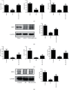
AKF-PD improved mitochondrial energy metabolism in the UUO and IRI models. (a) The expression of PDH, CS, and AKGDH in renal tissue from the indicated mice was measured by real-time PCR (n = 4). (b) The expression of CPT1 and ACOX1 in renal tissue from the indicated mice was determined by real-time PCR (n = 4). (c) The expression of complex I, complex II, and complex V in renal tissue from the indicated mice was analyzed by western blot (n = 4). (c) The expression of PDH, CS, and AKGDH in renal tissue from the indicated mice was examined by real-time PCR (n = 4). (d) The expression of complex I, complex II, and complex V in renal tissue from the indicated mice was measured by western blot (n = 4). P < 0.05 and ∗∗P < 0.01 UUO/IRI group vs. sham/control group; #P < 0.05 and ##P < 0.01 UUO+AKF-PD/IRI+AKF-PD group vs. UUO/IRI group. Data were analyzed by one-way ANOVA. AKF-PD: fluorofenidone; PDH: pyruvate dehydrogenase; CS: citrate synthase; AKGDH: α-ketoglutarate dehydrogenase; CPT1: carnitine palmitoyltransferase 1; ACOX1: acyl-CoA oxidase 1.
Figure 5
Figure 5
AKF-PD increased mitochondrial biogenesis in the UUO and IRI models. (a) The expression of PGC-1α, TFAM, and NRF1 in renal tissue from the indicated mice was measured by real-time PCR (n = 4). (b) The expression of SIRT1 in renal tissue from the indicated mice was measured by western blot (n = 4). (c) The expression of PGC-1α, TFAM, and NRF1 in renal tissue from the indicated mice was measured by real-time PCR (n = 4). (d) The expression of SIRT1 in renal tissue from the indicated mice was measured by western blot (n = 4). P < 0.05 and ∗∗P < 0.01 UUO/IRI group vs. sham/control group; #P < 0.05 and ##P < 0.01 UUO+AKF-PD/IRI+AKF-PD group vs. UUO/IRI group. Data were analyzed by one-way ANOVA. AKF-PD: fluorofenidone; PGC-1α: peroxisome proliferator-activated receptor γ coactivator-1α; TFAM: mitochondrial transcription factor A; NRF1: nuclear respiratory factor 1; SIRT1: sirtuin 1.
Figure 6
Figure 6
AKF-PD suppressed mitochondrial oxidative stress in the UUO model. (a) The expression of 4HNE in renal tissue from the indicated mice was determined by immunohistochemistry, ×200 (n = 4). (b) The expression of TRX2 and SOD2 in renal tissue from the indicated mouse group was measured by real-time PCR (n = 4). (c, d) The expression of NOX4, SIRT3, and SOD2 in renal tissue from the indicated mice was measured by western blot (n = 4). P < 0.05 and ∗∗P < 0.01 UUO group vs. sham group; #P < 0.05 and ##P < 0.01 UUO+AKF-PD group vs. UUO group. Data were analyzed by one-way ANOVA. AKF-PD: fluorofenidone; 4HNE: 4-hydroxynonenal; TRX2: thioredoxin 2; SOD2: superoxide dismutase 2; NOX4: NADPH oxidase 4; SIRT3: sirtuin 3.
Figure 7
Figure 7
AKF-PD suppressed mitochondrial oxidative stress in the IRI model. (a) The expression of 4HNE in renal tissue from the indicated mice was determined by immunohistochemistry, ×200 (n = 4). (b) The expression of TRX2 and SOD2 in renal tissue from the indicated mouse group was measured by real-time PCR (n = 4). (c, d) The expression of NOX4, SIRT3, and SOD2 in renal tissue from the indicated mice was measured by western blot (n = 4). P < 0.05 and ∗∗P < 0.01 IRI group vs. control group; #P < 0.05 and ##P < 0.01 IRI+AKF-PD group vs. IRI group. Data were analyzed by one-way ANOVA. AKF-PD: fluorofenidone; 4HNE: 4-hydroxynonenal; TRX2: thioredoxin 2; SOD2: superoxide dismutase 2; NOX4: NADPH oxidase 4; SIRT3: sirtuin 3.
Figure 8
Figure 8
AKF-PD reduced mitochondrial damage and mitochondrial oxidative stress in HK-2 cells. (a) The expression of E-cadherin and α-SMA in HK-2 cells was measured by western blot (n = 3). (b) Mitochondrial morphology of HK-2 cells was examined by transmission electron microscopy. (c) ATP levels of HK-2 cells were measured by the ATP assay kit (n = 3). (d) MitoSOX Deep Red fluorescence was used to detect mitochondrial ROS of HK-2 cells by flow cytometry analysis (n = 3). (e, f) The expression of SIRT3, SOD2, and NOX4 in HK-2 cells was measured by western blot (n = 3). P < 0.05 and ∗∗P < 0.01 TGF-β group vs. normal group; #P < 0.05 and ##P < 0.01 AKF-PD group vs. TGF-β group. Data were analyzed by one-way ANOVA. AKF-PD: fluorofenidone; N: normal group; TGF-β: transforming growth factor-β; α-SMA: alpha smooth muscle actin; SOD2: superoxide dismutase 2; NOX4: NADPH oxidase 4; SIRT3: sirtuin 3.

Similar articles

Cited by

References

    1. François H., Chatziantoniou C. Renal fibrosis: recent translational aspects. Matrix Biology . 2018;68:318–332. doi: 10.1016/j.matbio.2017.12.013. - DOI - PubMed
    1. Luo F., Xu R., Guanglai S., Xue D., He X., Ying X. Alleviation of TGF-β1 induced tubular epithelial-mesenchymal transition via the δ-opioid receptor. The FEBS Journal . 2021;288(4):1243–1258. doi: 10.1111/febs.15459. - DOI - PubMed
    1. Textor S. C., Lerman L. O. Paradigm shifts in atherosclerotic renovascular disease: where are we now? Journal of the American Society of Nephrology . 2015;26(9):2074–2080. doi: 10.1681/ASN.2014121274. - DOI - PMC - PubMed
    1. Mulay S. R., Chongxu S., Ma X., Joachim A. H. Novel insights into crystal-induced kidney injury. Kidney Diseases . 2018;4(2):49–57. doi: 10.1159/000487671. - DOI - PMC - PubMed
    1. Peng F., Li H., Li S., et al. Micheliolide ameliorates renal fibrosis by suppressing the Mtdh/BMP/MAPK pathway. Laboratory Investigation . 2019;99(8):1092–1106. doi: 10.1038/s41374-019-0245-6. - DOI - PMC - PubMed

Substances