. 2022 Mar 21:2022:8151917.
doi: 10.1155/2022/8151917. eCollection 2022.

Cysteine-Rich Whey Protein Isolate (CR-WPI) Ameliorates Erectile Dysfunction by Diminishing Oxidative Stress via DDAH/ADMA/NOS Pathway

Affiliations

Cysteine-Rich Whey Protein Isolate (CR-WPI) Ameliorates Erectile Dysfunction by Diminishing Oxidative Stress via DDAH/ADMA/NOS Pathway

Kefan Li et al. Oxid Med Cell Longev. .

Abstract

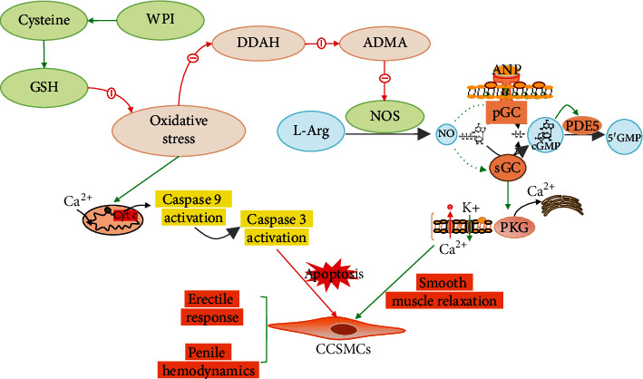
Nitric oxide synthase- (NOS-) dependent endothelial dysfunction induced by oxidative stress (OS) is assumed to play a pivotal role in the pathogenesis and progression of diabetes mellitus-related erectile dysfunction (DMED). Cysteine-rich whey protein isolate (CR-WPI) is a widely used protein supplement and has been confirmed to reduce reactive oxygen species (ROS) by increasing cellular antioxidant glutathione (GSH). However, it is currently unknown whether CR-WPI elicits therapeutic effects in DMED. Here, we provide diabetic rats with CR-WPI to determine its effect on DMED and the underlying mechanisms. The results suggest that CR-WPI supplementation increased GSH biosynthesis and reduced ROS content and simultaneously upregulated the dimethylarginine dimethylaminohydrolase (DDAH)/asymmetrical dimethylarginine (ADMA)/nitric oxide synthase (NOS) metabolic pathway. Evaluation of intracavernous pressure (ICP) also showed an improvement of penile erectile function in CR-WPI-treated rats. The results of the vitro cell culture showed that glutathione pretreatment protected corpus cavernosum smooth muscle cells (CCSMC) from H2O2-induced apoptosis by decreasing Caspase 9 and Caspase 3 expressions. These results augur well for the potential therapeutic application of dietary CR-WPI supplementation for treating diabetic erectile dysfunction.


Conflict of interest statement

Jimmy Gutman consults with a whey protein distributor.

Figures

Figure 1
Figure 1
Flow diagram of the animal experiment and erectile responses of rats after treatment. (a) The process of the establishment of DMED rat model and treatment of the experimental animal. (b) CR-WPI supplementation improves the erectile function of DMED rats. The red lines denote electrical stimulation of the cavernous nerve for 60 s. (c) The ratio of maximum ICP to MAP derived from all eight groups (n = 8) is presented as bar graphs, and the data are shown as mean ± standard deviation. (d) The total ICP is obtained by calculating the area under curve, and the data are shown as mean ± standard deviation. P < 0.05 and ∗∗P < 0.01 indicate a significant difference compared with the DM group. ##P < 0.01 indicates a significant difference compared with the sham group.
Figure 2
Figure 2
Effects of CR-WPI treatment on ROS, GSH, and ADMA levels in penile tissues after 30 days or 60 days treatment. (a) CR-WPI treatment increases GSH levels in the corpus cavernosum in a time and concentration-dependent manner. (b) CR-WPI treatment reduces ROS levels in diabetic rats. (c) CR-WPI supplementation decreases ADMA concentration of penile tissues in diabetic rats. P < 0.05 and ∗∗P < 0.01 indicate a significant difference compared with the DM group. ##P < 0.01 indicates a significant difference compared with the sham group.
Figure 3
Figure 3
Changes of the DDAH/NOS pathway and apoptosis-related proteins in penile tissues after 100 mg kg−1 of and 300 mg kg−1 CR-WPI supplementation for 60 days. (a) Representative immunofluorescence staining of the eNOS-positive penile specimen (red) in the sham, DM, 100 mg kg−1 CR-WPI, and 300 mg kg−1 CR-WPI groups for 60 days. (b) The effects of CR-WPI on the protein expression of DDAH, NOS, Caspase 3, and Caspase 9 in diabetic rats. β-Actin was used as a loading control. P < 0.01 and ∗∗P < 0.01 indicate a significant difference compared with the DM group. #P < 0.05 and ##P < 0.01 indicate a significant difference compared with the sham group.
Figure 4
Figure 4
CR-WPI treatment for 60 days increases the smooth muscle content of the corpus cavernosum. (a) Representative immunofluorescence staining of the α-SMA-positive corpus cavernosum positive for α-SMA in the sham, DM, 100 mg kg−1 CR-WPI, and 300 mg kg−1 CR-WPI groups. (b) Masson's trichrome staining of penile tissue in four groups after different treatments. Smooth muscle and collagen in the corpus cavernosum are stained red and blue, respectively. (c) The effect of CR-WPI on the ratio of smooth muscle to collagen and protein expression of α-SMA (ratio to β-actin) in the corpus cavernosum. Bars denote the mean densitometry ratio between smooth muscle content and collagen content per field. ∗∗P < 0.01 compared to the DM group and #P < 0.05 and ##P < 0.01 compared to the sham group.
Figure 5
Figure 5
Effect of GSH on CCSMC apoptosis. (a) Primary CCSMCs emerging from the corpus cavernosum tissue after 3 days. (b) Immunofluorescence with anti-α-SMA antibody for CCSMC identification, ×40 amplification. (c) Cell viability was detected by flow cytometry in the normal control group treated with PBS (NC) and the H2O2-treated groups (pretreated with 0, 5, and 10 mM GSH). (d) The average apoptotic cell percentages and expression of Caspase 3 and Caspase 9 of CCSMCs in the NC group, H2O2 group, 5 mM GSH, and 10 mM GSH group. Data are expressed as mean ± SD, n = 3. P < 0.05 and ∗∗P < 0.01 indicate a significant difference compared with the H2O2 group. #P < 0.05 and ##P < 0.01 indicate a significant difference compared with the NC group.
Figure 6
Figure 6
Diagram of the molecular mechanism of how CR-WPI affects erectile function. DDAH: dimethylarginine dimethylaminohydrolase; ADMA: asymmetric dimethylaminohydrolase; L-Arg: L-arginine; GC: guanylyl cyclase; PDE5: phosphodiesterase-5; PKG: cGMP-dependent protein kinase.

Similar articles

Cited by

References

    1. Ponholzer A., Temml C., Mock K., Marszalek M., Obermayr R., Madersbacher S. Prevalence and risk factors for erectile dysfunction in 2869 men using a validated questionnaire. European Urology . 2005;47(1):80–86. doi: 10.1016/j.eururo.2004.08.017. - DOI - PubMed
    1. Sáenz de Tejada I., Angulo J., Cellek S., et al. Pathophysiology of erectile dysfunction. The Journal of Sexual Medicine . 2005;2(1):26–39. doi: 10.1111/j.1743-6109.2005.20103.x. - DOI - PubMed
    1. Belba A., Cortelazzo A., Andrea G., et al. Erectile dysfunction and diabetes: association with the impairment of lipid metabolism and oxidative stress. Clinical Biochemistry . 2016;49(1-2):70–78. doi: 10.1016/j.clinbiochem.2015.10.004. - DOI - PubMed
    1. Tuncayengin A., Biri H., Onaran M., et al. Cavernosal tissue nitrite, nitrate, malondialdehyde and glutathione levels in diabetic and non-diabetic erectile dysfunction. International Journal of Andrology . 2003;26(4):250–254. doi: 10.1046/j.1365-2605.2003.00427.x. - DOI - PubMed
    1. Wortel R. C., Mizrachi A., Li H., et al. Sildenafil protects endothelial cells from radiation-induced oxidative stress. The Journal of Sexual Medicine . 2019;16(11):1721–1733. doi: 10.1016/j.jsxm.2019.08.015. - DOI - PMC - PubMed