

Chest X-ray findings in drug-sensitive and drug-resistant pulmonary tuberculosis patients in Uganda
- PMID: 35355939
- PMCID: PMC8958542
- DOI: 10.1016/j.jctube.2022.100312
Chest X-ray findings in drug-sensitive and drug-resistant pulmonary tuberculosis patients in Uganda
Abstract
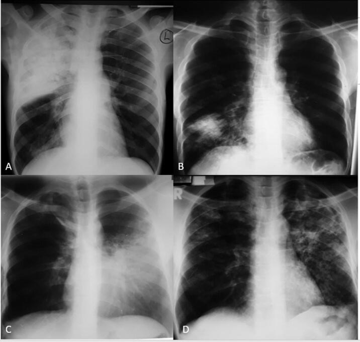
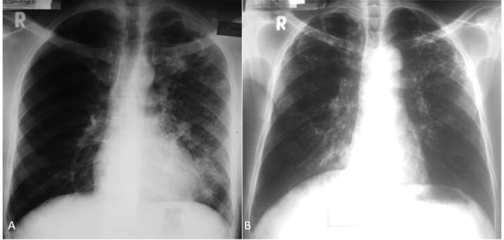
Background: Tuberculosis (TB) is one of the leading causes of death worldwide. Radiology has an important role in the diagnosis of both drug-sensitive (DS) and rifampicin-resistant (RR) pulmonary TB (PTB). This study aimed to compare the chest x-ray (CXR) patterns of microbiologically confirmed DS and RR PTB cases stratified by HIV serostatus in Uganda.
Methods: We conducted a hospital-based retrospective study at the Mulago National Referral Hospital (MNRH) TB wards. All participants had a microbiologically confirmed diagnosis of PTB. CXR findings extracted included infiltrates, consolidation, cavity, fibrosis, bronchiectasis, atelectasis, and other non-lung parenchymal findings. All films were examined by two independent radiologists blinded to the clinical diagnosis.
Results: We analyzed CXR findings of 165 participants: 139 DS- and 26 RR-TB cases. The majority (n = 118, 71.7%) of the participants were seronegative for HIV. Overall, 5/165 (3%) participants had normal CXR. There was no statistically significant difference in the proportion of participants with consolidations (74.8% versus 88.5%; p = 0.203), bronchopneumonic opacities (56.1% versus 42.3%, p = 0.207) and cavities (38.1% versus 46.2%, p = 0.514), across drug susceptibility status (DS versus RR TB). Among HIV-infected participants, consolidations were predominantly in the middle lung zone in the DS TB group and in the lower lung zone in the RR TB group (42.5% versus 12.8%, p = 0.66). HIV-infected participants with RR TB had statistically significantly larger cavity sizes compared to their HIV uninfected counterparts with RR TB (7.7 ± 6.8 cm versus 4.2 ± 1.3 cm, p = 0.004).
Conclusions: We observed that a vast majority of participants had similar CXR changes, irrespective of drug susceptibility status. However, HIV-infected RR PTB had larger cavities.The diagnostic utility of cavity sizes for the differentiation of HIV-infected and non-infected RR TB could be investigated further.
Keywords: Chest radiograph; DS-TB, Drug sensitive tuberculosis; Drug-sensitive; HIV, Human Immunodeficiency Virus; MDR, Multidrug resistant tuberculosis; MNRH, Mulago national referral hospital; MTB, Mycobacterium tuberculosis; PTB, Pulmonary Tuberculosis; Pulmonary tuberculosis; RIF, Resistance to rifampicin; RR-TB, Rifampicin-resistant tuberculosis; Rifampicin-resistant; WHO, World Health Organization.
© 2022 The Author(s).
Conflict of interest statement
The authors declare that they have no known competing financial interests or personal relationships that could have appeared to influence the work reported in this paper.
Figures
References
-
- World Health Organization. Global tuberculosis report 2020: executive summary [Internet]. Geneva: World Health Organization; 2020 [cited 2022 Mar 19]. 11 p. Available from: https://apps.who.int/iris/handle/10665/337538.
-
- Ubaidi BAA, Ubaidi BAA. The Radiological Diagnosis of Pulmonary Tuberculosis (TB) in Primary Care. [cited 2021 Aug 22]; Available from: https://clinmedjournals.org/articles/jfmdp/journal-of-family-medicine-an....
LinkOut - more resources
Full Text Sources
