Review
. 2022 Mar 9:9:861913.
doi: 10.3389/fcvm.2022.861913. eCollection 2022.

Clinical Imaging of the Penumbra in Ischemic Stroke: From the Concept to the Era of Mechanical Thrombectomy

Affiliations
Review

Clinical Imaging of the Penumbra in Ischemic Stroke: From the Concept to the Era of Mechanical Thrombectomy

Lucie Chalet et al. Front Cardiovasc Med. .

Abstract

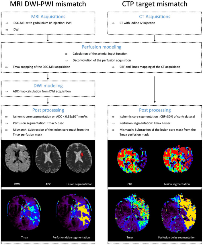
The ischemic penumbra is defined as the severely hypoperfused, functionally impaired, at-risk but not yet infarcted tissue that will be progressively recruited into the infarct core. Early reperfusion aims to save the ischemic penumbra by preventing infarct core expansion and is the mainstay of acute ischemic stroke therapy. Intravenous thrombolysis and mechanical thrombectomy for selected patients with large vessel occlusion has been shown to improve functional outcome. Given the varying speed of infarct core progression among individuals, a therapeutic window tailored to each patient has recently been proposed. Recent studies have demonstrated that reperfusion therapies are beneficial in patients with a persistent ischemic penumbra, beyond conventional time windows. As a result, mapping the penumbra has become crucial in emergency settings for guiding personalized therapy. The penumbra was first characterized as an area with a reduced cerebral blood flow, increased oxygen extraction fraction and preserved cerebral metabolic rate of oxygen using positron emission tomography (PET) with radiolabeled O2. Because this imaging method is not feasible in an acute clinical setting, the magnetic resonance imaging (MRI) mismatch between perfusion-weighted imaging and diffusion-weighted imaging, as well as computed tomography perfusion have been proposed as surrogate markers to identify the penumbra in acute ischemic stroke patients. Transversal studies comparing PET and MRI or using longitudinal assessment of a limited sample of patients have been used to define perfusion thresholds. However, in the era of mechanical thrombectomy, these thresholds are debatable. Using various MRI methods, the original penumbra definition has recently gained a significant interest. The aim of this review is to provide an overview of the evolution of the ischemic penumbra imaging methods, including their respective strengths and limitations, as well as to map the current intellectual structure of the field using bibliometric analysis and explore future directions.

Keywords: MRI; PET; cerebral metabolic rate of oxygen; ischemic thresholds; penumbra; thrombectomy; thrombolysis.


Conflict of interest statement

LC, DR, and TB are employees of Olea Medical, a company developing the OleaSphere platform including pipelines for ischemic penumbra. The remaining authors declare that the research was conducted in the absence of any commercial or financial relationships that could be construed as a potential conflict of interest.

Figures

Figure 1
Figure 1
Oxygen metabolism [15O]-Positron Emission Tomography (PET) imaging pipeline. PET images published in JNM. Heiss WD. Radionuclide imaging in ischemic stroke. J Nucl Med. (2014) 55:1831–41. © SNMMI. CMRO2, cerebral metabolic rate of oxygen; IDIF, image-derived input function; CaO2, arterial blood oxygen content; VOI, volume of interest; CBF, cerebral blood flow; OEF, oxygen extraction fraction; K1, tracer delivery rate.
Figure 2
Figure 2
Definitions of perfusion and metabolic penumbra from the gold standard [15O]-Positron Emission Tomography (PET) to the widely distributed Computed Tomography (CT) and Magnetic Resonance Imaging (MRI) perfusion imaging. PET images published in JNM. Heiss WD. Radionuclide imaging in ischemic stroke. J Nucl Med. (2014) 55:1831–41. SNMMI.; © CT and MRI images from Olea Medical. CBF, cerebral blood flow; CMRO2, cerebral metabolic rate of oxygen; OEF, oxygen extraction fraction; ADC, apparent diffusion coefficient; Tmax, time to maximum of residual function.
Figure 3
Figure 3
CT and MRI pipelines. In dark red: segmented infarct core. In yellow: segmented perfusion delay on Tmax. Images from Olea Medical. MRI, magnetic resonance imaging; DWI, diffusion weighted imaging; PWI, perfusion weighted imaging; CTP, computed tomography perfusion; DSC, dynamic susceptibility contrast; IV, intravenous bolus; CBF, cerebral blood flow; Tmax, time to maximum of residual function; ADC, apparent diffusion coefficient.
Figure 4
Figure 4
Summarized development timeline of the penumbra imaging field. Circles: the major contributions to the acute ischemic stroke research field. Arrows: the clinical validation years for reperfusion therapies. Rectangles: the major clinical trials using advanced imaging to extend therapeutic windows. Red and green code for mechanical thrombectomy and intravenous thrombolysis clinical trials, respectively. PET, positron emission tomography; MRI, magnetic resonance imaging; CT, computed tomography.
Figure 5
Figure 5
Co-citation network providing an overview of the acute ischemic stroke research field evolutions through its most contributing publications: Red cluster [1981–2005]: MRI diffusion-perfusion mismatch; Green cluster [2000–2013]: perfusion CT; Blue cluster [2003–2012]: Eligibility for thrombolysis; Yellow cluster [1995–2013]: Thrombolysis combined to endovascular therapies; Purple cluster [2015–2018]: Mechanical thrombectomy. MRI, magnetic resonance imaging; CT, computed tomography.
Figure 6
Figure 6
Bibliographic coupling network in the era of mechanical thrombectomy [2015–2021] with labeled clusters based on predominance of themes within the clusters. MRI, magnetic resonance imaging; CTP, computed tomography perfusion; MT, mechanical thrombectomy; APT, amide proton transfer; CEST, chemical exchange saturation transfer.
Figure 7
Figure 7
Detailed principle and pipeline for OEF mapping with the multiparametric quantitative Blood Oxygen Level Dependent (mqBOLD) method and CMRO2 mapping with streamlined qBOLD. MRI, magnetic resonance imaging; DSC, dynamic susceptibility contrast; IV, intravenous bolus; CBV, cerebral blood volume; CBF, cerebral blood flow.

References

    1. Wafa HA, Wolfe CDA, Emmett E, Roth GA, Johnson CO, Wang Y. Burden of stroke in Europe. Stroke. (2020) 51:2418–27. 10.1161/STROKEAHA.120.029606 - DOI - PMC - PubMed
    1. Ovbiagele B, Goldstein LB, Higashida RT, Howard VJ, Johnston SC, Khavjou OA, et al. . Forecasting the future of stroke in the United States: a policy statement from the American Heart Association and American Stroke Association. Stroke. (2013) 44:2361–75. 10.1161/STR.0b013e31829734f2 - DOI - PubMed
    1. Astrup J, Siesjo BK, Symon L. Thresholds in cerebral ischemia - the ischemic penumbra. Stroke. (1981) 12:723–5. 10.1161/01.STR.12.6.723 - DOI - PubMed
    1. Baron JC. Mapping the ischaemic penumbra with PET: implications for acute stroke treatment. Cerebrovasc Dis. (1999) 9:193–201. 10.1159/000015955 - DOI - PubMed
    1. Jones TH, Morawetz RB, Crowell RM, Marcoux FW, FitzGibbon SJ, DeGirolami U, et al. . Thresholds of focal cerebral ischemia in awake monkeys. J Neurosurg. (1981) 54:773–82. 10.3171/jns.1981.54.6.0773 - DOI - PubMed