. 2022 Mar 9:9:829546.
doi: 10.3389/fcvm.2022.829546. eCollection 2022.

Electrophysiologic Conservation of Epicardial Conduction Dynamics After Myocardial Infarction and Natural Heart Regeneration in Newborn Piglets

Affiliations

Electrophysiologic Conservation of Epicardial Conduction Dynamics After Myocardial Infarction and Natural Heart Regeneration in Newborn Piglets

Hanjay Wang et al. Front Cardiovasc Med. .

Abstract

Newborn mammals, including piglets, exhibit natural heart regeneration after myocardial infarction (MI) on postnatal day 1 (P1), but this ability is lost by postnatal day 7 (P7). The electrophysiologic properties of this naturally regenerated myocardium have not been examined. We hypothesized that epicardial conduction is preserved after P1 MI in piglets. Yorkshire-Landrace piglets underwent left anterior descending coronary artery ligation at age P1 (n = 6) or P7 (n = 7), After 7 weeks, cardiac magnetic resonance imaging was performed with late gadolinium enhancement for analysis of fibrosis. Epicardial conduction mapping was performed using custom 3D-printed high-resolution mapping arrays. Age- and weight-matched healthy pigs served as controls (n = 6). At the study endpoint, left ventricular (LV) ejection fraction was similar for controls and P1 pigs (46.4 ± 3.0% vs. 40.3 ± 4.9%, p = 0.132), but significantly depressed for P7 pigs (30.2 ± 6.6%, p < 0.001 vs. control). The percentage of LV myocardial volume consisting of fibrotic scar was 1.0 ± 0.4% in controls, 9.9 ± 4.4% in P1 pigs (p = 0.002 vs. control), and 17.3 ± 4.6% in P7 pigs (p < 0.001 vs. control, p = 0.007 vs. P1). Isochrone activation maps and apex activation time were similar between controls and P1 pigs (9.4 ± 1.6 vs. 7.8 ± 0.9 ms, p = 0.649), but significantly prolonged in P7 pigs (21.3 ± 5.1 ms, p < 0.001 vs. control, p < 0.001 vs. P1). Conduction velocity was similar between controls and P1 pigs (1.0 ± 0.2 vs. 1.1 ± 0.4 mm/ms, p = 0.852), but slower in P7 pigs (0.7 ± 0.2 mm/ms, p = 0.129 vs. control, p = 0.052 vs. P1). Overall, our data suggest that epicardial conduction dynamics are conserved in the setting of natural heart regeneration in piglets after P1 MI.

Keywords: conduction; electrophysiology; heart; mapping; myocardial infarction; neonate - age; regeneration.


Conflict of interest statement

The authors declare that the research was conducted in the absence of any commercial or financial relationships that could be construed as a potential conflict of interest.

Figures

Figure 1
Figure 1
Neonatal piglet myocardial infarction model and epicardial mapping. (A) Permanent ligation of the left anterior descending coronary artery (yellow arrow) was performed in piglets on postnatal day 1 or postnatal day 7 to induce myocardial infarction. The ischemic territory (below dotted yellow line) was visibly distinguished from the perfused myocardium by pallor and hypokinesis. (B,C) After 7 weeks, epicardial conduction mapping was performed using custom 3D-printed high-resolution mapping arrays with contoured shells matching the epicardial surface profile of the 7–8-week-old pig heart, ensuring optimal coverage and contact during mapping. Scale bar, 1 cm.
Figure 2
Figure 2
Piglets naturally recover left ventricular function after myocardial infarction on postnatal day 1. At 7 weeks after myocardial infarction (MI), cardiac magnetic resonance imaging was performed to evaluate left ventricular function, including (A) end-systolic volume (ESV) indexed against body surface area, (B) end-diastolic volume (EDV) indexed against body surface area, (C) stroke volume (SV) indexed against body surface area, (D) cardiac index, and (E) ejection fraction. Following heart explant, the (F) heart weight to body weight ratio was determined. No significant differences were observed between pigs receiving MI on postnatal day 1 (P1) compared to healthy controls. Pigs receiving MI on postnatal day 7 (P7), however, developed left ventricular dysfunction.
Figure 3
Figure 3
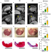
Piglets exhibit reduced scar formation after myocardial infarction on postnatal day 1. At 7 weeks after myocardial infarction (MI), cardiac magnetic resonance imaging (MRI) with late gadolinium enhancement was performed to evaluate left ventricular (LV) fibrosis. Representative long-axis (LAX) and short-axis (SAX) views are presented for (A) healthy controls, (B) pigs receiving MI on postnatal day 1 (P1), and (C) pigs receiving MI on postnatal day 7 (P7). Infarcted areas are marked by yellow arrows. Scale bars, 5 cm. (D) Compared to P7 pigs, P1 pigs developed significantly smaller infarcts, including (E) significantly smaller transmural infarcts. (F–H) Following heart explant, the LV endocardium was assessed for visible scar formation, revealing (I) significantly smaller endocardial scar in P1 pigs compared to P7 pigs. (J–L) Masson's trichome staining was performed to confirm the presence of fibrotic scar. Scale bars, 1 cm. (M) Significantly less fibrotic content was observed in P1 pigs compared to P7 pigs.
Figure 4
Figure 4
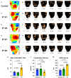
Piglets demonstrate conserved epicardial conduction dynamics after myocardial infarction on postnatal day 1. At 7 weeks after myocardial infarction (MI), epicardial conduction mapping was performed. Representative isochrone activation maps and timelapse activation maps are presented for (A,B) one healthy control, illustrating normal conduction through the right and left bundle branches and Purkinje system; (C–F) two pigs receiving MI on postnatal day 1 (P1) with similar activation patterns as healthy controls; and (G–J) two pigs receiving MI on postnatal day 7 (P7), including (G,H) one P7 pig which developed right bundle branch block, and (I,J) one P7 pig which developed left bundle branch block. (K) Apex activation time was similar between controls and P1 pigs, but profoundly delayed in P7 pigs. (L) Conduction velocity was similar between controls and P1 pigs, but slower in P7 pigs. (M) QRS interval was similar between controls and P1 pigs, but significantly longer in P7 pigs.

Similar articles

Cited by

References

    1. Roth GA, Mensah GA, Johnson CO, Addolorato G, Ammirati E, Baddour LM, et al. . Global Burden of Cardiovascular Diseases and Risk Factors, 1990-2019: Update From the GBD 2019 Study. J Am Coll Cardiol. (2020) 76:2982–3021. 10.1016/j.jacc.2020.11.010 - DOI - PMC - PubMed
    1. Ezekowitz JA, Kaul P, Bakal JA, Armstrong PW, Welsh RC, McAlister FA. Declining in-hospital mortality and increasing heart failure incidence in elderly patients with first myocardial infarction. J Am Coll Cardiol. (2009) 53:13–20. 10.1016/j.jacc.2008.08.067 - DOI - PubMed
    1. Bergmann O, Bhardwaj RD, Bernard S, Zdunek S, Barnabé-Heider F, Walsh S, et al. . Evidence for cardiomyocyte renewal in humans. Science. (2009) 324:98–102. 10.1126/science.1164680 - DOI - PMC - PubMed
    1. Elde S, Wang H, Woo YJ. The expanding armamentarium of innovative bioengineered strategies to augment cardiovascular repair and regeneration. Front Bioeng Biotechnol. (2021) 9:674172. 10.3389/fbioe.2021.674172 - DOI - PMC - PubMed
    1. Chen K, Huang Y, Singh R, Wang ZZ. Arrhythmogenic risks of stem cell replacement therapy for cardiovascular diseases. J Cell Physiol. (2020) 235:6257–67. 10.1002/jcp.29554 - DOI - PMC - PubMed