Microvascular and Prognostic Effect in Lesions With Different Stent Expansion During Primary PCI for STEMI: Insights From Coronary Physiology and Intravascular Ultrasound
- PMID: 35355977
- PMCID: PMC8959302
- DOI: 10.3389/fcvm.2022.816387
Microvascular and Prognostic Effect in Lesions With Different Stent Expansion During Primary PCI for STEMI: Insights From Coronary Physiology and Intravascular Ultrasound
Abstract
Background: While coronary stent implantation in ST-elevation myocardial infarction (STEMI) can mechanically revascularize culprit epicardial vessels, it might also cause distal embolization. The relationship between geometrical and functional results of stent expansion during the primary percutaneous coronary intervention (pPCI) is unclear.
Objective: We sought to determine the optimal stent expansion strategy in pPCI using novel angiography-based approaches including angiography-derived quantitative flow ratio (QFR)/microcirculatory resistance (MR) and intravascular ultrasound (IVUS).
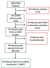
Methods: Post-hoc analysis was performed in patients with acute STEMI and high thrombus burden from our prior multicenter, prospective cohort study (ChiCTR1800019923). Patients aged 18 years or older with STEMI were eligible. IVUS imaging, QFR, and MR were performed during pPCI, while stent expansion was quantified on IVUS images. The patients were divided into three subgroups depending on the degree of stent expansion as follows: overexpansion (>100%), optimal expansion (80%-100%), and underexpansion (<80%). The patients were followed up for 12 months after PCI. The primary endpoint included sudden cardiac death, myocardial infarction, stroke, unexpected hospitalization or unplanned revascularization, and all-cause death.
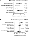
Results: A total of 87 patients were enrolled. The average stent expansion degree was 82% (in all patients), 117% (in overexpansion group), 88% (in optimal expansion), and 75% (in under-expansion). QFR, MR, and flow speed increased in all groups after stenting. The overall stent expansion did not affect the final QFR (p = 0.08) or MR (p = 0.09), but it reduced the final flow speed (-0.14 cm/s per 1%, p = 0.02). Under- and overexpansion did not affect final QFR (p = 0.17), MR (p = 0.16), and flow speed (p = 0.10). Multivariable Cox analysis showed that stent expansion was not the risk factor for MACE (hazard ratio, HR = 0.97, p = 0.13); however, stent expansion reduced the risk of MACE (HR = 0.95, p = 0.03) after excluding overexpansion patients. Overexpansion was an independent risk factor for no-reflow (HR = 1.27, p = 0.02) and MACE (HR = 1.45, p = 0.007). Subgroup analysis shows that mild underexpansion of 70%-80% was not a risk factor for MACE (HR = 1.11, p = 0.08) and no-reflow (HR = 1.4, p = 0.08); however, stent expansion <70% increased the risk of MACE (HR = 1.36, p = 0.04).
Conclusions: Stent expansion does not affect final QFR and MR, but it reduces flow speed in STEMI. Appropriate stent underexpansion of 70-80% does not seem to be associated with short-term prognosis, so it may be tolerable as noninferior compared with optimal expansion. Meanwhile, overexpansion and underexpansion of <70% should be avoided due to the independent risk of MACEs and no-reflow events.
Keywords: IVUS; ST-elevation myocardial infarction (STEMI); major adverse cardiovascular events (MACEs); microcirculation; stent expansion.
Copyright © 2022 Li, Sun, Luo, Yang, Ye, Guo, Xu, Sun, Zhang, Luo, Zhou, Tu and Dong.
Conflict of interest statement
The authors declare that the research was conducted in the absence of any commercial or financial relationships that could be construed as a potential conflict of interest.
Figures


References
-
- Fujii K, Carlier SG, Mintz GS, Yang YM, Moussa I, Weisz G, et al. Stent underexpansion and residual reference segment stenosis are related to stent thrombosis after sirolimus-eluting stent implantation: an intravascular ultrasound study. J Am Coll Cardiol. (2005) 45:995–8. 10.1016/j.jacc.2004.12.066 - DOI - PubMed
-
- De Maria GL, Cuculi F, Patel N, Dawkins S, Fahrni G, Kassimis G, et al. How does coronary stent implantation impact on the status of the microcirculation during primary percutaneous coronary intervention in patients with ST-elevation myocardial infarction? Eur Heart J. (2015) 36:3165–77. 10.1093/eurheartj/ehv353 - DOI - PMC - PubMed
-
- Safi H, Bourantas CV, Ramasamy A, Zanchin T, Bar S, Tufaro V, et al. Predictive value of the QFR in detecting vulnerable plaques in non-flow limiting lesions: a combined analysis of the PROSPECT and IBIS-4 study. Int J Cardiovasc Imaging. (2020) 36:993–1002. 10.1007/s10554-020-01805-9 - DOI - PubMed
LinkOut - more resources
Full Text Sources
Miscellaneous

