Vasa previa: A rare obstetric complication-A case series and a literature review
- PMID: 35356178
- PMCID: PMC8939036
- DOI: 10.1002/ccr3.5608
Vasa previa: A rare obstetric complication-A case series and a literature review
Abstract
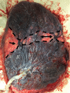
Vasa previa is a rare condition. However, since the increase in assisted reproductive technologies (ARTs), clinicians are more frequently confronted with this complication. In this study, we present five cases of vasa previa prenatally diagnosed from a tertiary referral hospital with approximately 2000 births yearly.
Keywords: assisted reproductive technology; insertio velamentosa; placenta; prenatal diagnosis; vasa previa.
© 2022 The Authors. Clinical Case Reports published by John Wiley & Sons Ltd.
Conflict of interest statement
The authors certify that they have no affiliations with or involvement in any organization or entity with any financial interest, or nonfinancial interest in the subject matter or materials discussed in this manuscript.
Figures


Similar articles
-
The Characteristics and Obstetric Outcomes of Type II Vasa Previa: Systematic Review and Meta-Analysis.Biomedicines. 2022 Dec 15;10(12):3263. doi: 10.3390/biomedicines10123263. Biomedicines. 2022. PMID: 36552018 Free PMC article. Review.
-
Current Evidence on Vasa Previa without Velamentous Cord Insertion or Placental Morphological Anomalies (Type III Vasa Previa): Systematic Review and Meta-Analysis.Biomedicines. 2023 Jan 7;11(1):152. doi: 10.3390/biomedicines11010152. Biomedicines. 2023. PMID: 36672661 Free PMC article. Review.
-
Impact of targeted scanning protocols on perinatal outcomes in pregnancies at risk of placenta accreta spectrum or vasa previa.Am J Obstet Gynecol. 2018 Apr;218(4):443.e1-443.e8. doi: 10.1016/j.ajog.2018.01.017. Epub 2018 Jan 17. Am J Obstet Gynecol. 2018. PMID: 29353034
-
Prenatally Diagnosed Vasa Previa: A Single-Institution Series of 96 Cases.Obstet Gynecol. 2016 Nov;128(5):1153-1161. doi: 10.1097/AOG.0000000000001680. Obstet Gynecol. 2016. PMID: 27741189
-
Clinical characteristics of a novel "Type 3" vasa previa: case series at a single center.J Matern Fetal Neonatal Med. 2022 Dec;35(25):7730-7736. doi: 10.1080/14767058.2021.1960975. Epub 2021 Aug 9. J Matern Fetal Neonatal Med. 2022. PMID: 34372741
Cited by
-
Core Outcome Set and Reporting Checklist for Studies on Vasa Previa.JAMA Netw Open. 2025 Mar 3;8(3):e251000. doi: 10.1001/jamanetworkopen.2025.1000. JAMA Netw Open. 2025. PMID: 40100217 Free PMC article.
-
A Case of Vasa Previa Diagnosed at Term: Elective Caesarean Section with Good Feto-Maternal Outcomes.Int Med Case Rep J. 2025 Jan 21;18:145-150. doi: 10.2147/IMCRJ.S459911. eCollection 2025. Int Med Case Rep J. 2025. PMID: 39866181 Free PMC article.
-
The Characteristics and Obstetric Outcomes of Type II Vasa Previa: Systematic Review and Meta-Analysis.Biomedicines. 2022 Dec 15;10(12):3263. doi: 10.3390/biomedicines10123263. Biomedicines. 2022. PMID: 36552018 Free PMC article. Review.
-
Current Evidence on Vasa Previa without Velamentous Cord Insertion or Placental Morphological Anomalies (Type III Vasa Previa): Systematic Review and Meta-Analysis.Biomedicines. 2023 Jan 7;11(1):152. doi: 10.3390/biomedicines11010152. Biomedicines. 2023. PMID: 36672661 Free PMC article. Review.
References
-
- Nohuz E, Boulay E, Gallot D, Lemery D, Vendittelli F. Can we perform a prenatal diagnosis of vasa previa to improve its obstetrical and neonatal outcomes? J Gynecol Obstet Hum Reprod. 2017;46(4):373‐377. - PubMed
-
- Lobstein . Arch de l’art des accouchments. Strasbourg. 1801;320‐28.
-
- Lubin B. Neonatal anaemia secondary to blood loss. Clin Haematol. 1978;7(1):19‐34. - PubMed
-
- Ruiter L, Kok N, Limpens J, et al. Incidence of and risk indicators for vasa praevia: a systematic review. BJOG. 2016;123(8):1278‐1287. - PubMed
-
- Robinson BK, Grobman WA. Effectiveness of timing strategies for delivery of individuals with vasa previa. Obstet Gynecol. 2011;117(3):542‐549. - PubMed
Publication types
LinkOut - more resources
Full Text Sources

