. 2022 Feb 1;6(1):69-89.
doi: 10.1162/netn_a_00210. eCollection 2022 Feb.

Disruption of large-scale electrophysiological networks in stroke patients with visuospatial neglect

Affiliations

Disruption of large-scale electrophysiological networks in stroke patients with visuospatial neglect

Tomas Ros et al. Netw Neurosci. .

Abstract

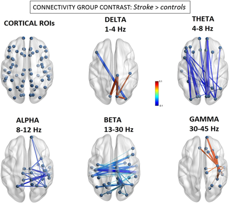
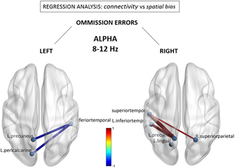
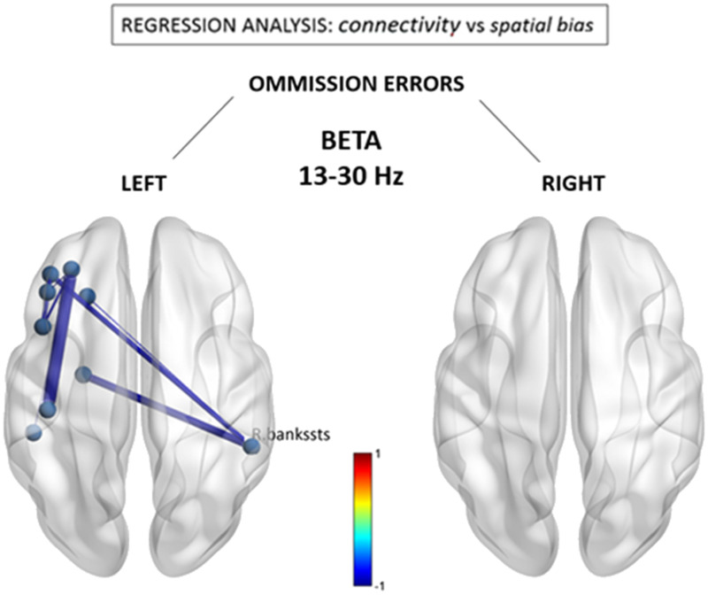
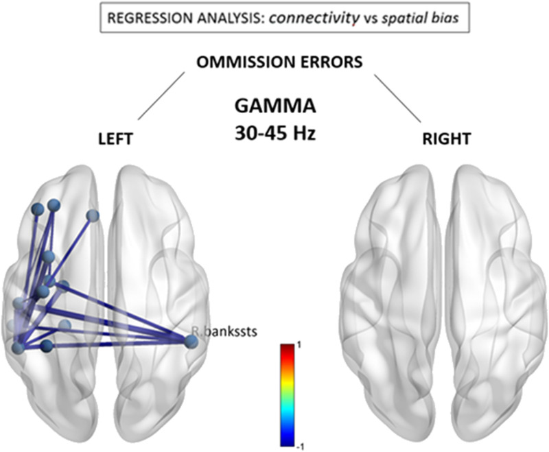
Stroke frequently produces attentional dysfunctions including symptoms of hemispatial neglect, which is characterized by a breakdown of awareness for the contralesional hemispace. Recent studies with functional MRI (fMRI) suggest that hemineglect patients display abnormal intra- and interhemispheric functional connectivity. However, since stroke is a vascular disorder and fMRI signals remain sensitive to nonneuronal (i.e., vascular) coupling, more direct demonstrations of neural network dysfunction in hemispatial neglect are warranted. Here, we utilize electroencephalogram (EEG) source imaging to uncover differences in resting-state network organization between patients with right hemispheric stroke (N = 15) and age-matched, healthy controls (N = 27), and determine the relationship between hemineglect symptoms and brain network organization. We estimated intra- and interregional differences in cortical communication by calculating the spectral power and amplitude envelope correlations of narrow-band EEG oscillations. We first observed focal frequency-slowing within the right posterior cortical regions, reflected in relative delta/theta power increases and alpha/beta/gamma decreases. Secondly, nodes within the right temporal and parietal cortex consistently displayed anomalous intra- and interhemispheric coupling, stronger in delta and gamma bands, and weaker in theta, alpha, and beta bands. Finally, a significant association was observed between the severity of left-hemispace search deficits (e.g., cancellation test omissions) and reduced functional connectivity within the alpha and beta bands. In sum, our novel results validate the hypothesis of large-scale cortical network disruption following stroke and reinforce the proposal that abnormal brain oscillations may be intimately involved in the pathophysiology of visuospatial neglect.

Keywords: Alpha; EEG; Functional network connectivity; Hemineglect; Stroke; sLORETA.


Figures

<b>Figure 1.</b>
Figure 1.
Group differences in EEG source functional connectivity. Significant differences of the magnitude of amplitude envelope correlations between stroke patients and controls. Line thickness represents larger absolute connectivity difference (Pearson’s correlation coefficient r). Red color indicates greater connectivity for patients versus controls, while blue color indicates greater connectivity for controls versus patients (only significant connectivity components are displayed, p < 0.05 network-based statistic corrected).
<b>Figure 2.</b>
Figure 2.
Alpha (8–12 Hz) connectivity as a function of neglect symptoms. Depicted connections correspond to changes in individual FNC components correlating with the number of omissions in the left and right hemifields, respectively, during the cancellation task. Red/blue values indicate statistically significant beta coefficients (p < 0.05 network-based statistic corrected). Further conjunction analysis (see text) revealed that reduced connections associated with left omissions (in left panel) were those showing the most significant overlap with general stroke-related decreases at the group level.
<b>Figure 3.</b>
Figure 3.
Beta (13–30 Hz) connectivity as a function of neglect symptoms. Depicted connections correspond to changes in individual FNC components correlating with the number of omissions in the left and right hemifields, respectively, during the cancellation task. Red/blue values indicate statistically significant beta coefficients (p < 0.05 network-based statistic corrected). R. bankssts = right bank of the superior temporal sulcus.
<b>Figure 4.</b>
Figure 4.
Gamma (30–40 Hz) connectivity as a function of neglect symptoms. Depicted connections correspond to changes in individual FNC components correlating with the number of omissions in the left and right hemifields, respectively, during the cancellation task. Red/blue values indicate statistically significant beta coefficients (p < 0.05 network-based statistic corrected). R.bankssts = right bank of the superior temporal sulcus.

References

    1. Akam, T. E., & Kullmann, D. M. (2012). Efficient “communication through coherence” requires oscillations structured to minimize interference between signals. PLoS Computational Biology, 8(11), e1002760. 10.1371/journal.pcbi.1002760, - DOI - PMC - PubMed
    1. Antzoulatos, E. G., & Miller, E. K. (2016). Synchronous beta rhythms of frontoparietal networks support only behaviorally relevant representations. ELife, 5. 10.7554/eLife.17822, - DOI - PMC - PubMed
    1. Baldassarre, A., Ramsey, L., Hacker, C. L., Callejas, A., Astafiev, S. V., Metcalf, N. V., … Corbetta, M. (2014). Large-scale changes in network interactions as a physiological signature of spatial neglect. Brain, 137, 3267–3283. 10.1093/brain/awu297, - DOI - PMC - PubMed
    1. Bauer, M., Kluge, C., Bach, D., Bradbury, D., Heinze, H. J., Dolan, R. J., & Driver, J. (2012). Cholinergic enhancement of visual attention and neural oscillations in the human brain. Current Biology, 22(5), 397–402. 10.1016/j.cub.2012.01.022, - DOI - PMC - PubMed
    1. Bentley, W. J., Li, J. M., Snyder, A. Z., Raichle, M. E., & Snyder, L. H. (2016). Oxygen level and LFP in task-positive and task-negative areas: Bridging BOLD fMRI and electrophysiology. Cerebral Cortex, 26(1), 346–357. 10.1093/cercor/bhu260, - DOI - PMC - PubMed

LinkOut - more resources