

miR-484: A Potential Biomarker in Health and Disease
- PMID: 35356223
- PMCID: PMC8959652
- DOI: 10.3389/fonc.2022.830420
miR-484: A Potential Biomarker in Health and Disease
Abstract
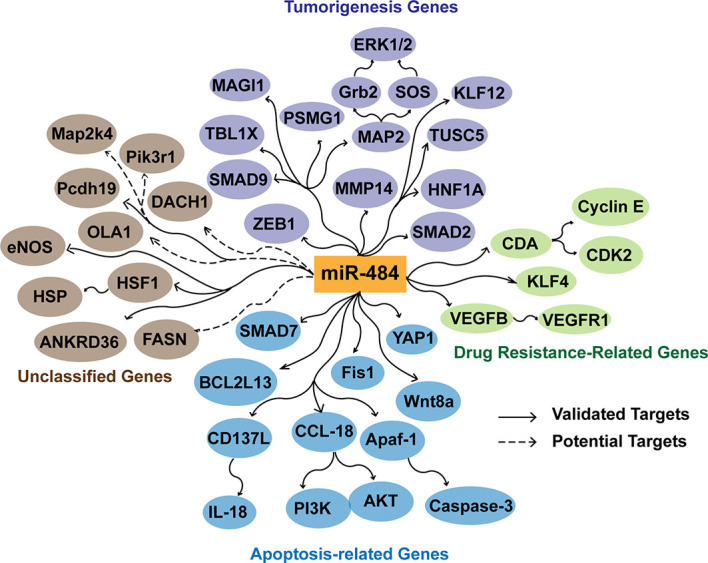
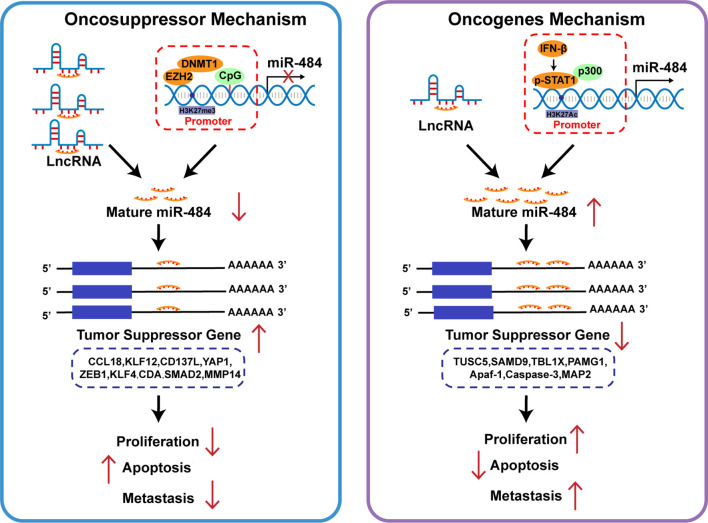
Disorders of miR-484 expression are observed in cancer, different diseases or pathological states. There is accumulating evidence that miR-484 plays an essential role in the development as well as the regression of different diseases, and miR-484 has been reported as a key regulator of common cancer and non-cancer diseases. The miR-484 targets that have effects on inflammation, apoptosis and mitochondrial function include SMAD7, Fis1, YAP1 and BCL2L13. For cancer, identified targets include VEGFB, VEGFR2, MAP2, MMP14, HNF1A, TUSC5 and KLF12. The effects of miR-484 on these targets have been documented separately. Moreover, miR-484 is typically described as an oncosuppressor, but this claim is simplistic and one-sided. This review will combine relevant basic and clinical studies to find that miR-484 promotes tumorigenesis and metastasis in liver, prostate and lung tissues. It will provide a basis for the possible mechanisms of miR-484 in early tumor diagnosis, prognosis determination, disease assessment, and as a potential therapeutic target for tumors.
Keywords: apoptosis; cancer; metastasis; physiological conditions; proliferation.
Copyright © 2022 Jia, Liu, Wang and Song.
Conflict of interest statement
The authors declare that the research was conducted in the absence of any commercial or financial relationships that could be construed as a potential conflict of interest.
Figures
Similar articles
-
DNMT1 recruited by EZH2-mediated silencing of miR-484 contributes to the malignancy of cervical cancer cells through MMP14 and HNF1A.Clin Epigenetics. 2019 Dec 7;11(1):186. doi: 10.1186/s13148-019-0786-y. Clin Epigenetics. 2019. PMID: 31810492 Free PMC article.
-
LncRNA NNT-AS1 promotes non-small cell lung cancer progression through regulating miR-22-3p/YAP1 axis.Thorac Cancer. 2020 Mar;11(3):549-560. doi: 10.1111/1759-7714.13280. Epub 2020 Jan 10. Thorac Cancer. 2020. PMID: 31923353 Free PMC article.
-
miR-375 induces docetaxel resistance in prostate cancer by targeting SEC23A and YAP1.Mol Cancer. 2016 Nov 10;15(1):70. doi: 10.1186/s12943-016-0556-9. Mol Cancer. 2016. PMID: 27832783 Free PMC article.
-
Dysregulated expression of miR‑367 in disease development and its prospects as a therapeutic target and diagnostic biomarker (Review).Biomed Rep. 2023 Oct 10;19(6):91. doi: 10.3892/br.2023.1673. eCollection 2023 Dec. Biomed Rep. 2023. PMID: 37901877 Free PMC article. Review.
-
miR-30d-5p: A Non-Coding RNA With Potential Diagnostic, Prognostic and Therapeutic Applications.Front Cell Dev Biol. 2022 Jan 27;10:829435. doi: 10.3389/fcell.2022.829435. eCollection 2022. Front Cell Dev Biol. 2022. PMID: 35155437 Free PMC article. Review.
Cited by
-
miR-484 as an "OncomiR" in Breast Cancer Promotes Tumorigenesis by Suppressing Apoptosis Genes.Ann Surg Oncol. 2025 Apr;32(4):2994-3008. doi: 10.1245/s10434-024-16656-0. Epub 2024 Dec 18. Ann Surg Oncol. 2025. PMID: 39692982
-
Exploring miRNA‑target gene profiles associated with drug resistance in patients with breast cancer receiving neoadjuvant chemotherapy.Oncol Lett. 2024 Feb 16;27(4):158. doi: 10.3892/ol.2024.14291. eCollection 2024 Apr. Oncol Lett. 2024. PMID: 38426156 Free PMC article.
-
Ceramide Metabolism Regulated by Sphingomyelin Synthase 2 Is Associated with Acquisition of Chemoresistance via Exosomes in Human Leukemia Cells.Int J Mol Sci. 2022 Sep 13;23(18):10648. doi: 10.3390/ijms231810648. Int J Mol Sci. 2022. PMID: 36142562 Free PMC article.
-
Non-coding RNAs in hepatocellular carcinoma: Insights into regulatory mechanisms, clinical significance, and therapeutic potential.Front Immunol. 2022 Oct 10;13:985815. doi: 10.3389/fimmu.2022.985815. eCollection 2022. Front Immunol. 2022. PMID: 36300115 Free PMC article. Review.
-
Whole Transcriptome Analysis of Intervention Effect of Sophora subprostrate Polysaccharide on Inflammation in PCV2 Infected Murine Splenic Lymphocytes.Curr Issues Mol Biol. 2023 Jul 20;45(7):6067-6084. doi: 10.3390/cimb45070383. Curr Issues Mol Biol. 2023. PMID: 37504299 Free PMC article.
References
Publication types
LinkOut - more resources
Full Text Sources