. 2022 Mar 14:28:1610217.
doi: 10.3389/pore.2022.1610217. eCollection 2022.

Correlations Between the Expression of Stromal Cell Activation Related Biomarkers, L-NGFR, Phospho-ERK1-2 and CXCL12, and Primary Myelofibrosis Progression

Affiliations

Correlations Between the Expression of Stromal Cell Activation Related Biomarkers, L-NGFR, Phospho-ERK1-2 and CXCL12, and Primary Myelofibrosis Progression

Tamas Szekely et al. Pathol Oncol Res. .

Abstract

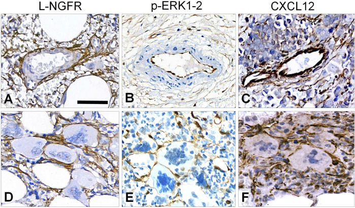
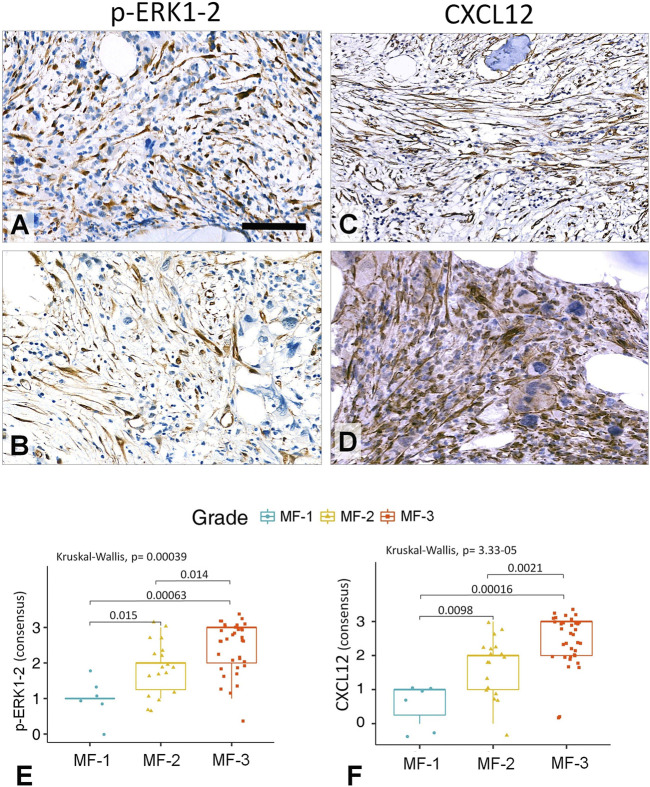
In myelofibrosis, pathologically enhanced extracellular matrix production due to aberrant cytokine signalling and clonal megakaryocyte functions result(s) in impaired hemopoiesis. Disease progression is still determined by detecting reticulin and collagen fibrosis with Gomori's silver impregnation. Here, we tested whether the expression growth related biomarkers L-NGFR/CD271, phospho-ERK1-2 and CXCL12 can be linked to the functional activation of bone marrow stromal cells during primary myelofibrosis progression. Immunoscores for all tested biomarkers showed varying strength of positive statistical correlation with the silver impregnation based myelofibrosis grades. The intimate relationship between spindle shaped stromal cells positive for all three markers and aberrant megakaryocytes was likely to reflect their functional cooperation. L-NGFR reaction was restricted to bone marrow stromal cells and revealed the whole length of their processes. Also, L-NGFR positive cells showed the most intersections, the best statistical correlations with myelofibrosis grades and the strongest interrater agreements. CXCL12 reaction highlighted stromal cell bodies and a weak extracellular staining in line with its constitutive release. Phospho-ERK1-2 reaction showed a similar pattern to CXCL12 in stromal cells with an additional nuclear staining in agreement with its role as a transcription factor. Both p-ERK1-2 and CXCL12 were also expressed at a moderate level in sinus endothelial cells. Connexin 43 gap junction communication channels, known to be required for CXCL12 release to maintain stem cell niche, were also expressed progressively in the myelofibrotic stromal network as a support of compartmental functions. Our results suggest that, diverse growth related pathways are activated in the functionally coupled bone marrow stromal cells during myelofibrosis progression. L-NGFR expression can be a useful biological marker of stromal cell activation which deserves diagnostic consideration for complementing Gomori's silver impregnation.

Keywords: -CXCL12; -L-NGFR/CD271; -connexin 43 channels; -phospho-ERK1-2; -stromal cell activation; primary myelofibrosis progression.


Conflict of interest statement

The authors declare that this study received shared facilities from 3DHISTEC Ltd. AC has been both the employee of 3DHISTECH Ltd. and a PhD student at Semmelweis University under the Co-operative PhD Program funded by the Hungarian Ministry of Innovation. TK is Member of the Editorial Board regularly reviewing manuscripts for POR. TS is a PhD student at Semmelweis University which supports PhD students publications in POR. The remaining authors declare that the research was conducted in the absence of any commercial or financial relationships that could be construed as a potential conflict of interest.

Figures

FIGURE 1
FIGURE 1
Staining pattern of the tested immunoreactions in intact areas of MF-1 bone marrow samples compared to Gomori’s silver impregnation (A). L-NGFR highlights random stromal cells with their thin processes (B). Phosphorylated ERK1-2 (p-ERK1-2) is detected mainly in oval nuclei of stromal and endothelial cells (C), while CXCL12 reaction stains cell bodies of these elements (D) with both revealing only few cell processes. DAB immunoperoxidase reactions (brown) counterstained using hematoxylin (B–D). Scale bar: 50 µm.
FIGURE 2
FIGURE 2
Immunoreaction patterns of the tested markers in arteries, periarterial stroma and abnormal megakaryocytes in myelofibrosis. L-NGFR positive stromal network spread from adventitial pericytes without endothelial reaction (A); while both p-ERK1-2 (B) and CXCL12 (C) reactions occur in endothelial and inter-arteriolar stromal cells with rare interconnections (upper row). Immunopositive elongated cell processes are intimately associated with aberrant megakaryocytes with all three markers (D, E and F; lower panel). DAB immunoperoxidase reactions (brown) counterstained using hematoxylin. Scale bar is 100 µm on A and B; and 50 µm on C-F.
FIGURE 3
FIGURE 3
Statistical correlations between L-NGFR immunoreaction scores and Gomori’s silver staining based myelofibrosis grades as recognized by the individual assessors after using both the Kruskal-Wallis test and the Wilcoxon post-hoc test (A–C). Pearson’s correlation values among scores of L-NGFR immunoreactions revealed between pairs of assessors in relation to Gomori’s silver staining based myelofibrosis grades (D–F).
FIGURE 4
FIGURE 4
Correlations between the L-NGFR immunopositive stromal network (A–C) and the reticulin (and collagen) scaffolding (D–F) in myelofibrotic bone marrow samples. The gradually increasing density with some interconnections among L-NGFR positive cell processes show good correlation with reticular (and collagen) fibrosis determining myelofibrosis grade as revealed by Gomori’s silver impregnation. DAB immunoperoxidase reactions (brown) counterstained using hematoxylin (A–C). Scale bar: 50 µm. High statistical correlation was detected between the consensus scores of the three assessors and the sliver impregnation based fibrosis grades both with the Kruskal-Wallis global trend test and the pairwise Wilcoxon post-hoc test (G).
FIGURE 5
FIGURE 5
Representative examples of grade 3 myelofibrotic bone marrow samples where either p-ERK1-2 (A,B) or CXCL12 (C,D) immunoreactions reveal elongated stromal cells, which however, are rarely interconnected by their processes into contiguous meshworks. DAB immunoperoxidase reactions (brown) counterstained using hematoxylin. Scale bar is 50 µm on A, B and D; and 100 µm on C. Both the p-ERK1-2 (E) and CXCL12 (F) immunoreaction consensus scores showed statistical correlations with reticulin-silver staining based myelofibrosis grades either when using Kruskal-Wallis trend test or the pairwise Wilcoxon test. However, their discrimination power was weaker than that of L-NGFR.
FIGURE 6
FIGURE 6
Double immunofluorescence for L-NGFR (green, A) and Cx43 (red, B) along with Hoechst nuclear counterstain (blue) in a grade 3 myelofibrosis. Merged images proved massive colocalization of the biomarkers (C), which became apparent after highlighting the green (in turquoise) and red signals (merged in yellow) with the image segmentation algorithm of the HistoQuant software (D). Semiautomated image analysis of the area fractions of fluorescent signals with this program in 67 standard areas of 10 myelofibrotic marrow samples (E) revealed strong overlap and correlation between L-NGFR and Cx43 signals suggesting that most particulate Cx43 belong to L-NGFR positive stromal cells (F). Scale bar on A is 25 µm on A-D and 500 µm on E.

Similar articles

Cited by

References

    1. Barbui T, Thiele J, Gisslinger H, Kvasnicka HM, Vannucchi AM, Guglielmelli P, et al. The 2016 WHO Classification and Diagnostic Criteria for Myeloproliferative Neoplasms: Document Summary and In-Depth Discussion. Blood Cancer J (2018) 8:15. 10.1038/s41408-018-0054-y - DOI - PMC - PubMed
    1. Tefferi A. Primary Myelofibrosis: 2019 Update on Diagnosis, Risk-Stratification and Management. Am J Hematol (2018) 93:1551–60. 10.1002/ajh.25230 - DOI - PubMed
    1. Schieber M, Crispino JD, Stein B. Myelofibrosis in 2019: Moving beyond JAK2 Inhibition. Blood Cancer J (2019) 9:74. 10.1038/s41408-019-0236-2 - DOI - PMC - PubMed
    1. Gömöri G. Silver Impregnation of Reticulum in Paraffin Sections. Am J Pathol (1937) 13:993–5. - PMC - PubMed
    1. Kvasnicka HM, Beham‐Schmid C, Bob R, Dirnhofer S, Hussein K, Kreipe H, et al. Problems and Pitfalls in Grading of Bone Marrow Fibrosis, Collagen Deposition and Osteosclerosis - a Consensus‐based Study. Histopathology (2016) 68:905–15. 10.1111/his.12871 - DOI - PubMed

MeSH terms