. 2022 May;29(3):e12744.
doi: 10.1111/xen.12744. Epub 2022 Mar 31.

Progressive genetic modifications of porcine cardiac xenografts extend survival to 9 months

Affiliations

Progressive genetic modifications of porcine cardiac xenografts extend survival to 9 months

Muhammad M Mohiuddin et al. Xenotransplantation. 2022 May.

Abstract

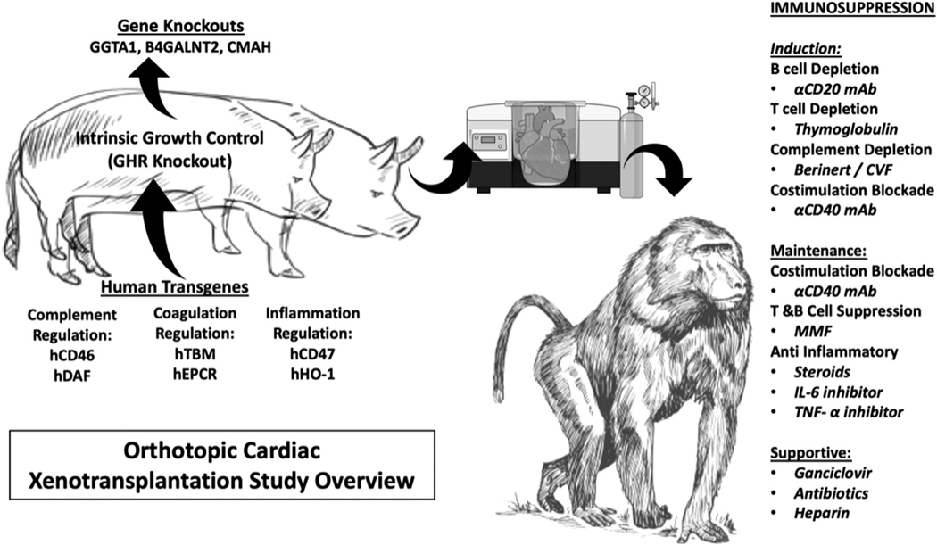
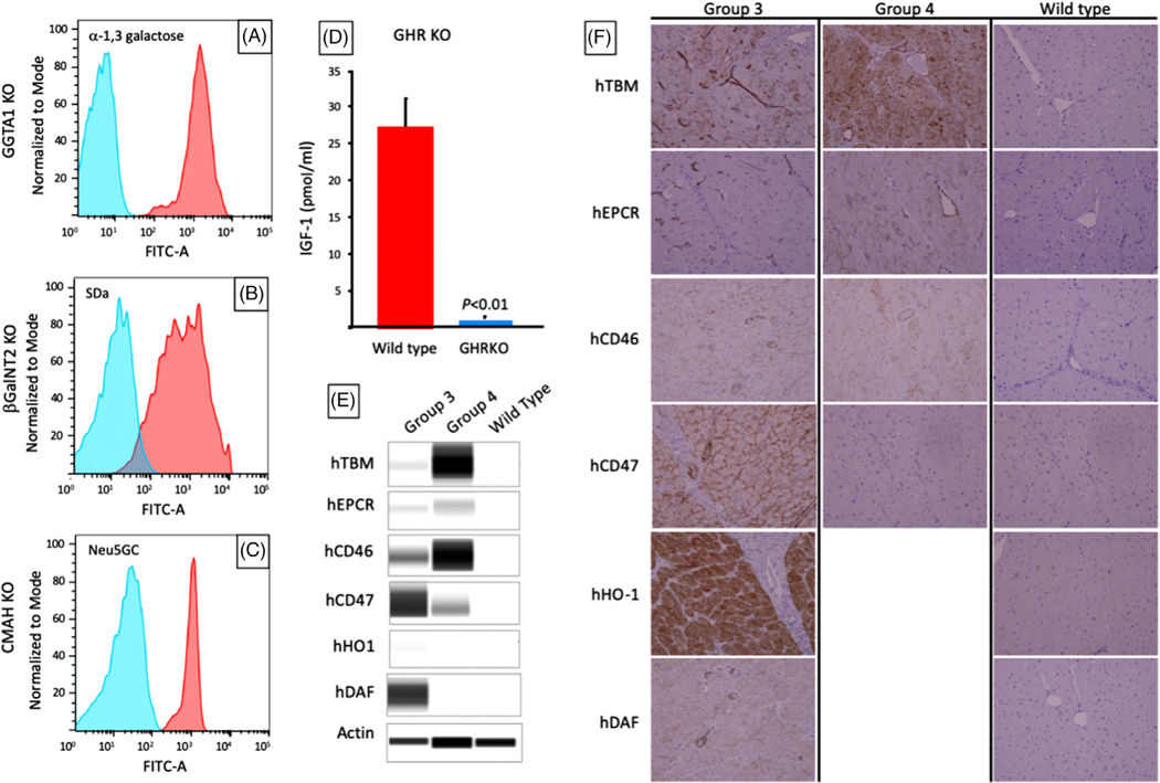
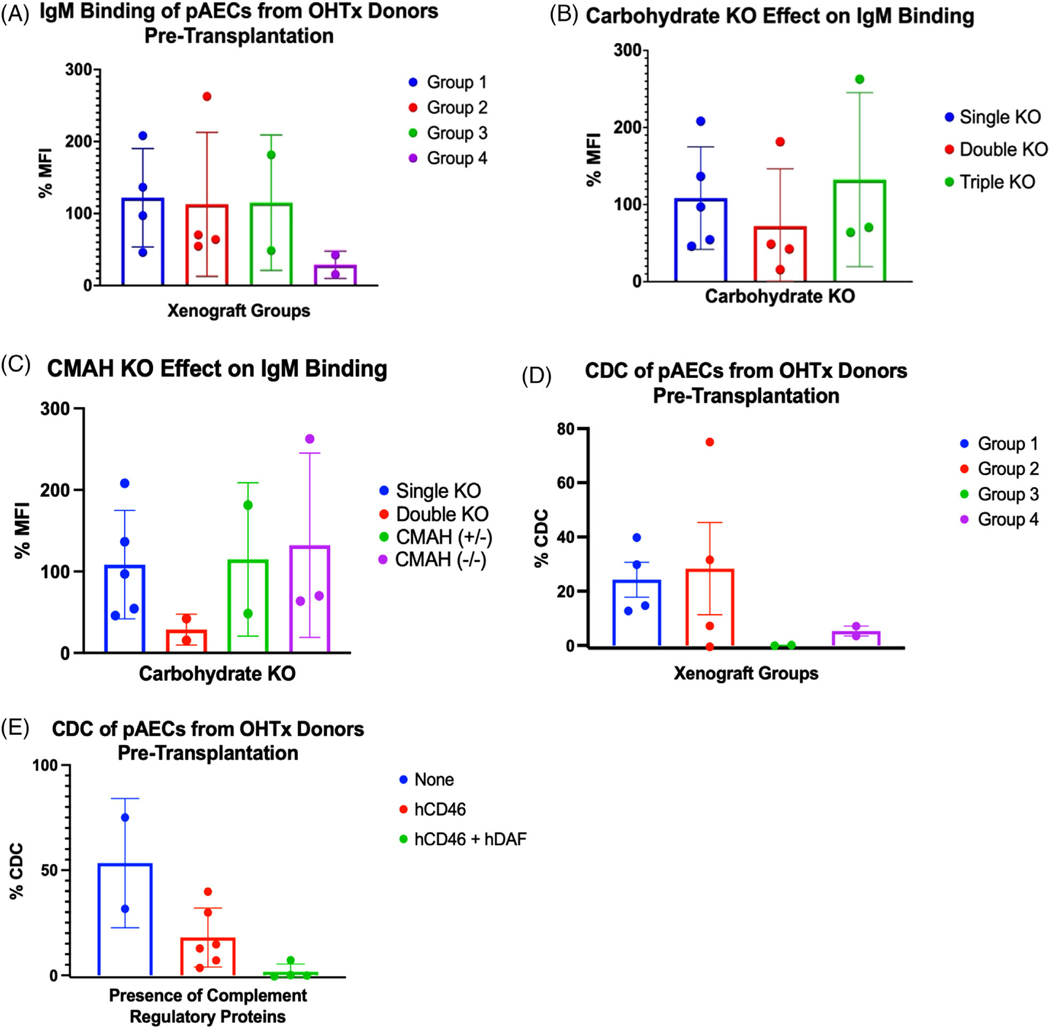
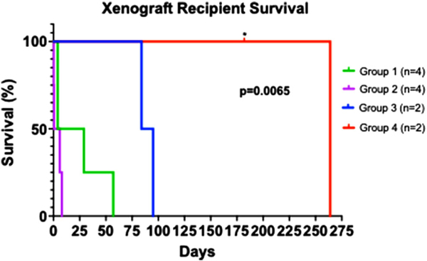
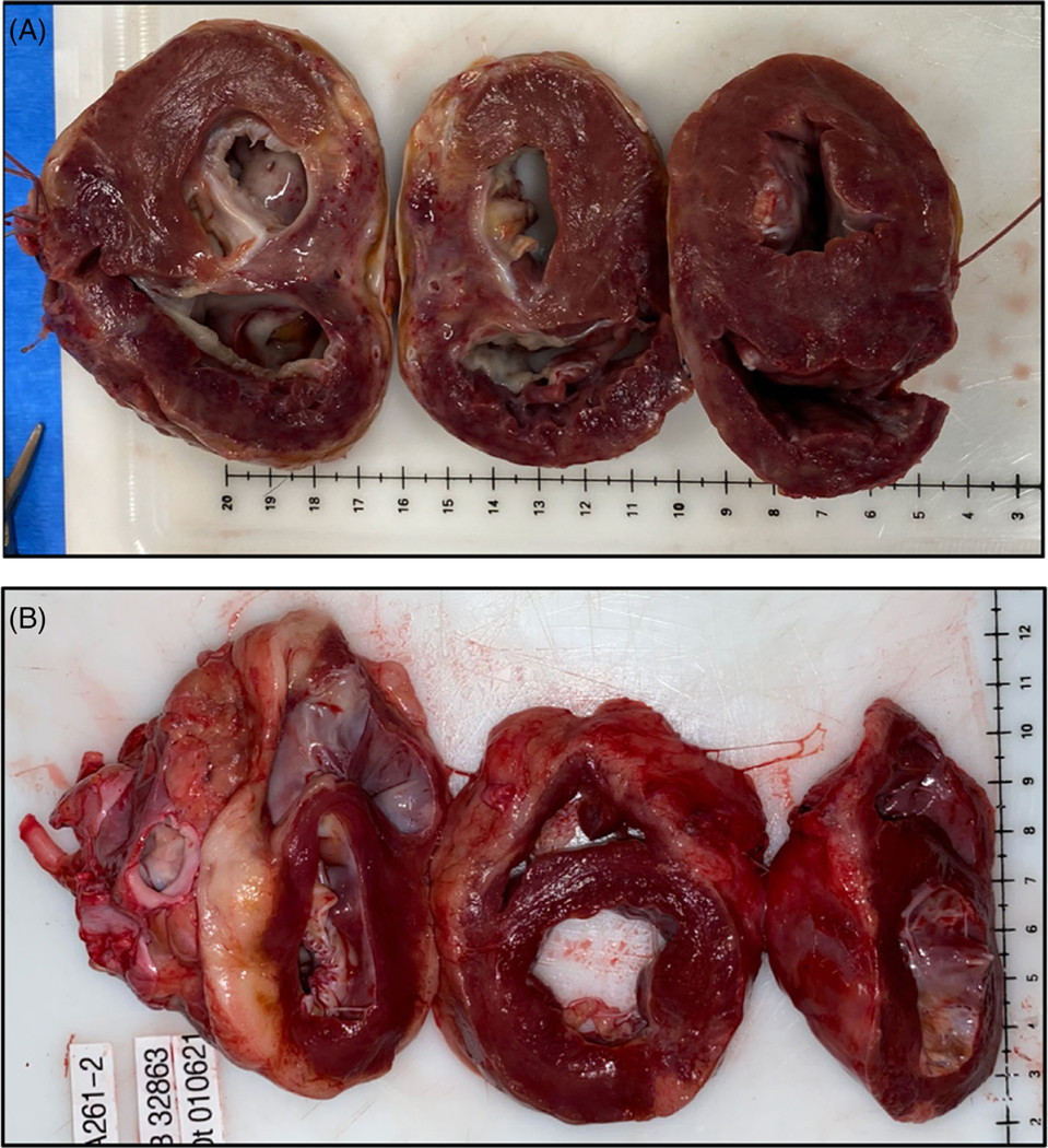
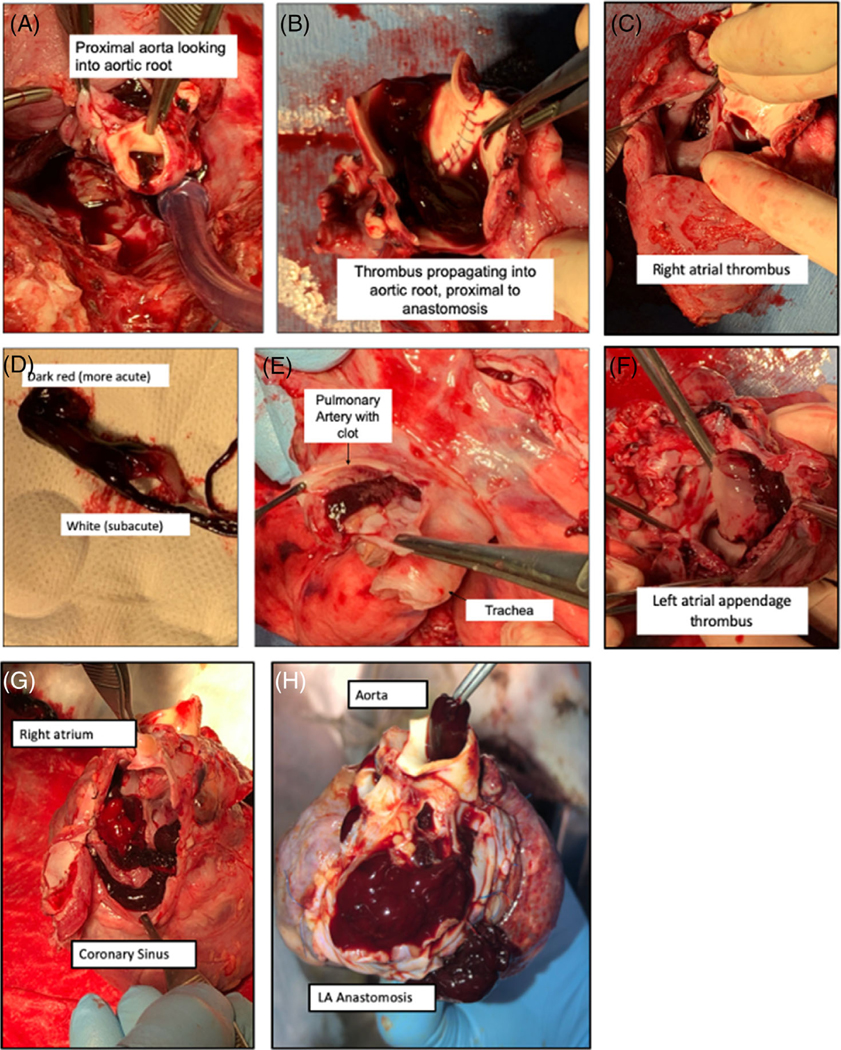
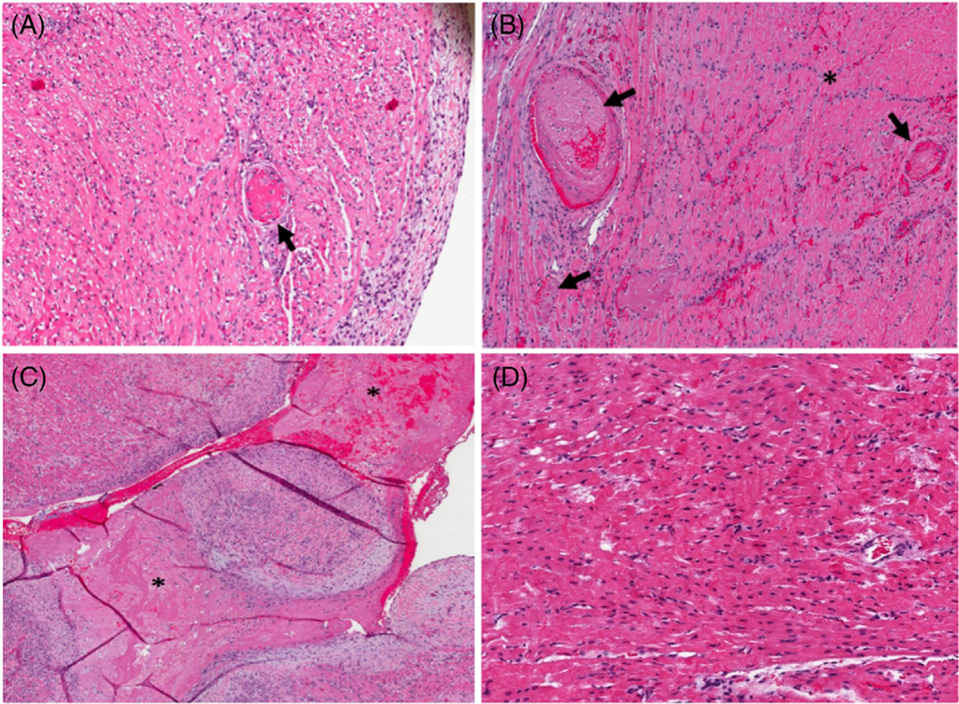
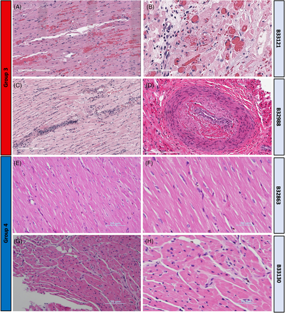
We report orthotopic (life-supporting) survival of genetically engineered porcine cardiac xenografts (with six gene modifications) for almost 9 months in baboon recipients. This work builds on our previously reported heterotopic cardiac xenograft (three gene modifications) survival up to 945 days with an anti-CD40 monoclonal antibody-based immunosuppression. In this current study, life-supporting xenografts containing multiple human complement regulatory, thromboregulatory, and anti-inflammatory proteins, in addition to growth hormone receptor knockout (KO) and carbohydrate antigen KOs, were transplanted in the baboons. Selective "multi-gene" xenografts demonstrate survival greater than 8 months without the requirement of adjunctive medications and without evidence of abnormal xenograft thickness or rejection. These data demonstrate that selective "multi-gene" modifications improve cardiac xenograft survival significantly and may be foundational for paving the way to bridge transplantation in humans.

Keywords: CRISPR; cardiac xenotransplantation; heart failure; pig heart; xenotransplantation.


Conflict of interest statement

CONFLICT OF INTEREST

David Ayares, Will Eyestone, Amy Dandro, Kasinath Kuravi, Lori Sorrels, and Todd Vaught are employed by Revivicor/United Therapeutics. The remaining authors declare that the research was conducted in the absence of any commercial or financial relationships that could be construed as a potential conflict of interest.

Figures

FIGURE 1
FIGURE 1. Study overview.
Pig-to-baboon xenotransplantation was performed with genetically modified pigs of various combinations. An anti-CD40 mAb-based regimen was used, and xenograft survival was measured. After euthanasia, the graft was explanted and examined. Multimodal analyses were performed on both the graft and the recipient
FIGURE 2
FIGURE 2. Xenograft phenotypes in long-term survivors.
(A–C) Flow cytometry showing the absence of a-1,3 galactose, SDa, and Neu5Gc antigens after knockout of GGTA1, B4GalNT2, and CMP-N-acetylneuraminic acid hydroxylase (CMAH), respectively in Group 2 pigs. Knockouts are shown in blue, wild types in red. CMAHKO (−/−) is shown in blue, whereas CMAHKO (+/−) has similar staining to wild type (not shown). (D) Serum IGF-1 levels in GHR knockout donors in Group 4 (blue) versus wild type pigs (red); (E) western blot of human transgenes expression in tail biopsies of Group 3 and 4 donor pigs. (F) Immunohistochemistry (IHC) of explanted heart xenografts from Group 3 and 4 donors showing expression of human transgenes (x200)
FIGURE 3
FIGURE 3. Characterization of multi-gene cardiac xenografts.
(A) IgM binding of pAECs from either xenograft donors or donor litter mates exposed to serum from Groups 1–4 recipients prior to OHTx. (B) IgM binding from panel a, grouped by single, double or triple knockout (KO) xenografts. (C) IgM binding from (A) and (B), grouped by CMP-N-acetylneuraminic acid hydroxylase (CMAH) (+/−) versus (−/−). Single = GGTA1KO, double = GGTA1KO and B4GalNT2, triple = GGTA1KO, B4GalNT2, and CMAHKO. %MFI = MFI as a percent of control. Complement dependent cytotoxicity (CDC) and IgM binding were performed as triplicates and presented here as an average of triplicates. (D) CDC measured on pig aortic endothelial cells (pAECs) from either xenograft donors or donor litter mates exposed to serum from Group 1–4 recipients prior to orthotopic transplantation (OHTx). (E) CDC from panel (D), grouped by complement regulatory proteins hCD46 and hDAF
FIGURE 4
FIGURE 4. Recipient survival of Groups 1–4.
Survival defined as time after transplantation before requiring euthanasia for deteriorating condition. * = death censored for euthanasia required. All other grafts contained histologic evidence of cardiac abnormalities contributing to deterioration requiring euthanasia. p = .0065 by Log-rank (Mantel-cox) test, suggesting a significant difference in survival between Groups 1–4
FIGURE 5 |
FIGURE 5 |. GHRKO versus non-GHRKO xenografts.
(A) non-GHRKO grafts (Group 3) exhibited biventricular wall thickening. Here, B33121 survived 84 days prior to requiring euthanasia for symptoms of diastolic heart failure. (B) GHRKO graft (Group 4) exhibiting normal histology without thickening at 182 days post-transplantation. This animal (B32863) was euthanized for weight loss as required by our institutional animal care committee but was exhibiting excellent graft function
FIGURE 6a
FIGURE 6a. (A) Thrombotic complications in Group 2 (xenografts without thromboregulatory proteins).
panels (A–F), showing B33167’s xenograft at explantation. Consists of propagating thrombus of the aortic root (A, B, and D), left and right atrial thrombus (C and F) and pulmonary artery (E). Pulmonary artery and left atrium appear to have acute and subacute components. Intracardiac thrombosis of B33156 within coronary sinus (G), aorta, and pulmonary vasculature (H).
FIGURE 6b
FIGURE 6b. (B) Histologic findings on Hematoxylin and eosin (H&E) in Group 2 (xenografts without thromboregulatory proteins)
(A) B33167 right ventricle, 10× magnification. Fibrin thrombus (arrow) in a background of ischemic myocytes. (B) B33156 apex, 10× magnification. Fibrin thrombi (arrows) and a region of ischemic myocytes (asterisk). (C) B33156 left ventricle, 10× magnification. Note the intracardiac organizing thrombus (asterisk). (D) B33060 right ventricle, 10×, note contraction bands and hypereosinophilia, indicating an early necrotic process
FIGURE 7
FIGURE 7. H&E in long-term survivors between Groups 3 and 4.
(A) Group 3, B33121 LV (17X)- congestion, mild interstitial hemorrhage individual myofiber degeneration and necrosis. (B) Group 3, B33121 right ventricle (RV) (40X) veins with intravascular thrombosis. (C) Group 3, B32988 LV (14X) interstitial mononuclear lymphoplasmacytic inflammation, scant perivascular hemorrhage, myodegeneration. (D) Group 3, B32988 RV (20X) organized thrombus in muscular artery, consistent with chronic xenograft vasculopathy. (E) Group 4, B32863 RV (20X) normal myocardium without evidence of rejection. (F) Group 4, B32863 RV (20X) normal myocardium without evidence of rejection. (G) Group 4, B33130 RV (20X), endomyocardial biopsy on POD#220, normal myocardium without evidence of rejection. (H) Group 4, B33130 RV (40X), endomyocardial biopsy, normal myocardium without evidence of rejection
FIGURE 8
FIGURE 8. IHC quantification and microthrombi.
Presented as averages within each group. High power field was with 20x magnification, resulting in approximately an 870 micron field. Microthrombi were graded on a scale of 0–4+, where 1+≥ 0 capillaries stained; 2+≥ 1–5 capillaries stained; 3+≥ 5–10 capillaries stained, 4+≥ 10 capillaries stained, per high power field
FIGURE 9
FIGURE 9. CD20+ cells in long-term survivors.
B-cell depletion is adequate after induction therapy, followed by reconstitution by 60–90 days after transplantation. Percent CD20+ cells calculated as a proportion of total CD3+ cells. Group 3 = B32988 and B33121; Group 4 = B32863 and B33130
FIGURE 10
FIGURE 10. Potential mechanisms of posttransplantation cardiac growth in xenotransplantation.
Posttransplantation cardiac xenograft growth is likely caused by both intrinsic and extrinsic factors, which includes rejection, intrinsic factors such as native xenograft growth. Other potential causes of growth include extrinsic factors such as physiologic mismatch leading to adaptive hypertrophy but were not observed in this study

Similar articles

Cited by

References

    1. Mohiuddin MM, Singh AK, Corcoran PC, et al. Chimeric 2C10R4 anti-CD40 antibody therapy is critical for long-term survival of GTKO.hCD46.hTBM pig-to-primate cardiac xenograft. Nat Commun. 2016;7:11138. - PMC - PubMed
    1. Iwase H, Ekser B, Satyananda V, et al. Pig-to-baboon heterotopic heart transplantation - exploratory preliminary experience with pigs transgenic for human thrombomodulin and comparison of three costimulation blockade-based regimens. Xenotransplantation. 2015;22:211–220. - PMC - PubMed
    1. Längin M, Mayr T, Reichart B, et al. Consistent success in life-supporting porcine cardiac xenotransplantation. Nature. 2018;564:430–433. - PubMed
    1. Shah JA, Patel MS, Elias N, et al. Prolonged survival following pig-toprimate liver xenotransplantation utilizing exogenous coagulation factors and costimulation blockade. Am J Transplant Off J Am Soc Transplant Am Soc Transpl Surg. 2017;17:2178–2185. - PMC - PubMed
    1. Yamamoto T, Hara H, Foote J, et al. Life-supporting kidney xenotransplantation from genetically engineered pigs in baboons: a comparison of two immunosuppressive regimens. Transplantation. 2019;103:2090–2104. - PubMed

Publication types

Substances