. 2022 Apr 27;10(2):e0270421.
doi: 10.1128/spectrum.02704-21. Epub 2022 Mar 31.

The cwp66 Gene Affects Cell Adhesion, Stress Tolerance, and Antibiotic Resistance in Clostridioides difficile

Affiliations

The cwp66 Gene Affects Cell Adhesion, Stress Tolerance, and Antibiotic Resistance in Clostridioides difficile

Qingshuai Zhou et al. Microbiol Spectr. .

Abstract

Clostridioides difficile is a Gram-positive, spore-forming anaerobic bacteria that is one of the leading causes of antibiotic-associated diarrhea. The cell wall protein 66 gene (cwp66) encodes a cell wall protein, which is the second major cell surface antigen of C. difficile. Although immunological approaches, such as antibodies and purified recombinant proteins, have been implemented to study the role of Cwp66 in cell adhesion, no deletion mutant of the cwp66 gene has yet been characterized. We constructed a cwp66 gene deletion mutant using Clustered Regularly Interspaced Short Palindromic Repeats Cpf1 (CRISPR-Cpf1) system. The phenotypic and transcriptomic changes of the Δcwp66 mutant compared with the wild-type (WT) strain were studied. The deletion of the cwp66 gene led to the decrease of cell adhesive capacity, cell motility, and stresses tolerance (to Triton X-100, acidic environment, and oxidative stress). Interestingly, the Δcwp66 mutant is more sensitive than the WT strain to clindamycin, ampicillin, and erythromycin but more resistant than the latter to vancomycin and metronidazole. Moreover, mannitol utilization capability in the Δcwp66 mutant was lost. Comparative transcriptomic analyses indicated that (i) 22.90-fold upregulation of cwpV gene and unable to express gpr gene were prominent in the Δcwp66 mutant; (ii) the cwp66 gene was involved in vancomycin resistance of C. difficile by influencing the expression of d-Alanine-d-Alanine ligase; and (iii) the mannose/fructose/sorbose IIC and IID components were upregulated in Δcwp66 mutant. The present work deepens our understanding of the contribution of the cwp66 gene to cell adhesion, stress tolerance, antibiotic resistance, and mannitol transportation of C. difficile. IMPORTANCE The cell wall protein 66 gene (cwp66) encodes a cell wall protein, which is the second major cell surface antigen of C. difficile. Although immunological approaches, such as antibodies and purified recombinant proteins, have been implemented to study the role of Cwp66 in cell adhesion, no deletion mutant of the cwp66 gene has yet been characterized. The current study provides direct evidence that the cwp66 gene serves as a major adhesion in C. difficile, and also suggested that deletion of the cwp66 gene led to the decrease of cell adhesive capacity, cell motility, and stresses tolerance (to Triton X-100, acidic environment, and oxidative stress). Interestingly, the antibiotic resistance and carbon source utilization profiles of the Δcwp66 mutant were significantly changed. These phenotypes were detrimental to the survival and pathogenesis of C. difficile in the human gut and may shed light on preventing C. difficile infection.

Keywords: CRISPR-Cpf1; Clostridioides difficile; cell wall protein 66 (Cwp66); phenotypic analysis; transcriptome analysis.


Conflict of interest statement

The authors declare no conflict of interest.

Figures

FIG 1
FIG 1
Verification of Δcwp66 gene mutant. (A) Gene context of the cwp66 gene, the cwp66 gene is flanked by CD630_27880 and CD630_27900 genes. (B) The primary structure of cwp66 gene, it consists of a signal peptide (SP), three cell wall binding 2 domains (CWB2), and a variable domain. (C) Verification of Δcwp66 mutant by using diagnostic PCR. Lane 1, PCR amplicon using genomic DNA of Δcwp66 mutant as the template; Lane 2, PCR amplicon using genomic DNA of WT strain as the template; lane M, DNA marker (from the top to the bottom, 10 k, 8 k, 6 k, 5 k, 4 k, 3 k, 2 k, 1.5 k, 1 k, 0.75 k, 0.5 k and 0.2 k). The reduction of PCR products indicates that the coding sequence of the cwp66 gene was deleted from the C. difficile 630 genome. (D) Verification of Δcwp66 mutant by using SDS-PAGE. Lane 1, cell lysate of the Δcwp66 mutant; Lane 2, cell lysate of the WT strain. The results showed that the Δcwp66 mutant strain absented a 66 kDa protein band than the WT strain (red arrows); lane M molecular weight marker (from the top to the bottom, 245 kDa, 180 kDa, 135 kDa, 100 kDa, 75 kDa, 63 kDa and 48 kDa). (E) Verification of Δcwp66 mutant Western blot analysis using the Cwp66 protein-specific antibodies. Lane 1, cell lysate of the WT strain; Lane 2, cell lysate of the Δcwp66 mutant; Lane 3, cell lysate of the ΔPaLoc strain, in which the pathogenicity locus (PaLoc) of C. difficile 630 strain was deleted, and the cwp66 gene in the ΔPaLoc strain was intact; Lane M, molecular weight marker (from the top to the bottom, 95 kDa, 72 kDa, 55 kDa, 43 kDa, 34 kDa and 26 kDa). The red arrowhead indicates the absent the Cwp66 band.
FIG 2
FIG 2
Changes in cells surface morphology of C. difficile strains. Cells surface morphology of the WT strain at ×5, 000 (A), ×10, 000 (B), ×20, 000 (C), and ×30, 000 (D) magnification. The first view of cells surface morphology of the Δcwp66 strain at ×5, 000 (E), ×10, 000 (F), ×20, 000 (G), and ×30, 000 (H) magnification (red arrows indicate cell disruption on the cell surface, green arrows indicate filamentous structure). The second view of cells surfaces morphology of the Δcwp66 strain at ×5, 000 (I), ×10, 000 (J), ×20, 000 (K), and ×30, 000 (L) magnification (red arrows indicate cell disruption on the cell surface, green arrows indicate filamentous structure). Cells surface morphology of the ::cwp66 strain at ×5, 000 (M), ×10, 000 (N), ×20, 000 (O) and ×30, 000 (P) magnification.
FIG 3
FIG 3
Phenotypic analysis of the Δcwp66 mutant. (A) Growth curves of WT, Δcwp66 and ::cwp66 mutants, the horizontal coordinate is incubation time (hours), the vertical coordinate is cell turbidity (OD600). (B) Cell autolysis rate of WT, Δcwp66 and ::cwp66 mutants after treated with Triton X-100, the horizontal coordinate is treatment duration of Triton X-100, the vertical coordinate is the percentage of unautolysed cells, the “-CT” suffix means untreated control. (C) Changes of cell tolerance to peroxide, the horizontal coordinate is the concentration of H2O2, the vertical coordinate is the cell turbidity (OD600). (D) Changes in cell adhesion ability of WT, Δcwp66, and ::cwp66 mutants under anaerobic condition, the horizontal coordinate indicates different strains, the vertical coordinate indicates the CFU counts of cells adhered to Caco-2 cells. The adhesion ability of Δcwp66 was decreased significantly (decreased more than 3-fold compared with the WT strain), and the ::cwp66 mutant restored 83% of adhesion ability compared with the WT strain. (E) Comparison of motility of the WT, Δcwp66, and ::cwp66 mutants, the motility of the Δcwp66 mutant strain was slightly decreased than that of the WT and the ::cwp66 stain restored cell motility. (F) Growth of WT, Δcwp66, and ::cwp66 mutants on BHI plate at pH = 4 and 5. The Δcwp66 strain failed to grow in BHI solid medium at pH = 4. In comparison, the WT and ::cwp66 strains grew well on the BHI medium at pH = 4. Student’s t test was used to compare the differences between groups, and the results were expressed as mean ± standard deviation, with a test level of α = 0.05. **, P < 0.01; ***, P < 0.001.
FIG 4
FIG 4
Changes of resistance profiles to antibiotics of the Δcwp66 mutant. The vertical coordinate is the value of OD600 and the horizontal coordinate is the antibiotic concentration (μg/mL). The red, light blue, and light green bars indicate the OD600 values of WT, Δcwp66, and ::cwp66 strain at different antibiotic concentrations. The Δcwp66 mutant is more sensitive than the WT strain to clindamycin, ampicillin, and erythromycin but more resistant than the latter to vancomycin and metronidazole. Except for chloramphenicol (due to plasmid born chloramphenicol transacetylase gene), overexpression of the cwp66 gene in the Δcwp66 mutant restored antibiotics resistant profiles for all tested antibiotics. Student’s t test was used to compare the differences between groups, and the results were expressed as mean ± standard deviation (n = 3), with a test level of α = 0.05. n, P > 0.1; ., P > 0.05; *, P < 0.05; **, P < 0.01; ***, P < 0.001.
FIG 5
FIG 5
Gene ontology classification. The horizontal coordinate is the term of GO level 2, the vertical coordinate is the –log10(P-value) enriched for each term, and the number on the column is the number of differential genes enriched to the corresponding term.
FIG 6
FIG 6
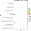
The GO enrichment analysis. The 20 most significantly enriched GO Term entries are displayed. The vertical coordinate is the GO term entries; the horizontal coordinate is the Rich factor, and the size of the bubbles indicates the number of differential genes enriched to the term, and the color indicates the false discovery rate (FDR) value of the pathway.
FIG 7
FIG 7
Annotation result against the KEGG database. The horizontal coordinate is the pathway's name; the vertical coordinate is the –log10 (P-value) enrichment for each pathway, and the number on the column is the number of differential genes enriched to the corresponding term.
FIG 8
FIG 8
The GO enrichment analysis. The 16 most significantly enriched KEGG pathways are displayed. The vertical coordinate is the KEGG pathway; the horizontal coordinate is the rich factor, and the size of the bubbles indicates the number of differential genes enriched to the pathway, and the color indicates the FDR value of the pathway.

References

    1. Lawson PA, Citron DM, Tyrrell KL, Finegold SM. 2016. Reclassification of Clostridium difficile as Clostridioides difficile (Hall and O’Toole 1935) Prévot 1938. Anaerobe 40:95–99. doi:10.1016/j.anaerobe.2016.06.008. - DOI - PubMed
    1. Pantaléon V, Soavelomandroso AP, Bouttier S, Briandet R, Roxas B, Chu M, Collignon A, Janoir C, Vedantam G, Candela T. 2015. The Clostridium difficile protease Cwp84 modulates both biofilm formation and cell-surface properties. PLoS One 10:e0124971. doi:10.1371/journal.pone.0124971. - DOI - PMC - PubMed
    1. Wen X, Shen C, Xia J, Zhong L-L, Wu Z, Ahmed M-G-S, Long N, Ma F, Zhang G, Wu W, Luo J, Xia Y, Dai M, Zhang L, Liao K, Feng S, Chen C, Chen Y, Luo W, Tian G-B. 2022. Whole-genome sequencing reveals the high nosocomial transmission and antimicrobial resistance of Clostridioides difficile in a single center in China, a four-year retrospective study. Microbiol Spectr 10:e0132221. doi:10.1128/spectrum.01322-21. - DOI - PMC - PubMed
    1. Péchiné S, Denève-Larrazet C, Collignon A. 2016. Clostridium difficile Adhesins, p 91–101. In Roberts AP, Mullany P (ed), Clostridium difficile. Springer, New York, NY. doi:10.1007/978-1-4939-6361-4_7. - DOI - PubMed
    1. Navarre WW, Schneewind O. 1999. Surface proteins of gram-positive bacteria and mechanisms of their targeting to the cell wall envelope. Microbiol Mol Biol Rev 63:174–229. doi:10.1128/MMBR.63.1.174-229.1999. - DOI - PMC - PubMed

Publication types

LinkOut - more resources