. 2022 Jun 6;221(5):e202202030.
doi: 10.1083/jcb.202202030. Epub 2022 Mar 31.

Structural and biochemical insights into lipid transport by VPS13 proteins

Affiliations

Structural and biochemical insights into lipid transport by VPS13 proteins

Jyoti Adlakha et al. J Cell Biol. .

Abstract

VPS13 proteins are proposed to function at contact sites between organelles as bridges for lipids to move directionally and in bulk between organellar membranes. VPS13s are anchored between membranes via interactions with receptors, including both peripheral and integral membrane proteins. Here we present the crystal structure of VPS13s adaptor binding domain (VAB) complexed with a Pro-X-Pro peptide recognition motif present in one such receptor, the integral membrane protein Mcp1p, and show biochemically that other Pro-X-Pro motifs bind the VAB in the same site. We further demonstrate that Mcp1p and another integral membrane protein that interacts directly with human VPS13A, XK, are scramblases. This finding supports an emerging paradigm of a partnership between bulk lipid transport proteins and scramblases. Scramblases can re-equilibrate lipids between membrane leaflets as lipids are removed from or inserted into the cytosolic leaflet of donor and acceptor organelles, respectively, in the course of protein-mediated transport.


Figures

Figure 1.
Figure 1.
Architecture of the VAB and its Pro-X-Pro motif binding site. (A) Schematic showing domain architecture of Vps13p. Residue numbers refer to the C. thermophilum Vps13p sequence. (B) Ribbons diagram for the VAB from C. thermophilum, showing its six repeated modules. The Pro-X-Pro motif is colored magenta. Inset shows one module, colored from blue at the N-terminus to red at the C-terminus, and a topology diagram colored in the same way. Fig. S1 A shows differences between the crystal structure and the AlphaFold2 prediction. (C) Sequence conservation, based on an alignment of 1,000 fungal Vps13s as determined by Consurf (Ashkenazy et al., 2016) and mapped onto the VAB surface. A patch centered on the sixth repeat and including the interface between the fifth and sixth repeats, outlined in yellow, is highly conserved and is the binding site for the receptor Pro-X-Pro motif. For electrostatic potential mapping, see Fig. S1 B. (D) Difference density from a 2Fo-Fc map (3.0 I/σ contour level), into which the Pro-X-Pro binding motif was modeled, is shown. Top: Two views of the Pro-X-Pro binding motif (yellow) bound to the VAB (light blue). Residues in the VAB binding surface, including those that were mutated to abrogate binding of the Pro-X-Pro motif, are labeled (mutated residues underlined). The asparagine in the sixth module at the end of β1, which is conserved in all modules, and which was mutated in previous interaction studies (Dziurdzik et al., 2020), is labeled (N2521). In the protein interior, it is part of an extensive hydrogen bonding network that stabilizes module folding (Fig. S1 C).
Figure S1.
Figure S1.
Details of the VAB structure. (A) Comparison of PXP-VAB1–6 in the crystal structure with the prediction from AlphaFold2. AlphaFold2 accurately predicted the fold of the individual modules as well as the interfaces between modules 1 + 2 and 2 + 3. The remaining interfaces in the crystal structure differed from those in the AlphaFold2 model. At left, the structures are superimposed based on the first three modules. The positions/orientations of the remaining modules differ in the two structures. In the box, the structures are superimposed based on the sixth module only. The interface between modules 5 + 6, where the Pro-X-Pro motif binds, is different in the two models. The AlphaFold2 model does not feature the groove that is the Pro-X-Pro motif binding site. (B) The electrostatic potential as calculated by APBS software (Jurrus et al., 2018) mapped onto the surface of the VAB. The VAB is shown in the same orientations as in Fig. 1 C. (C) The asparagine at the end of β-strand 1, strictly conserved in all repeat modules, is involved in an extensive hydrogen bonding network that stabilizes folding of the module.
Figure 2.
Figure 2.
The Pro-X-Pro motifs of Mcp1p, Spo71p, and Ypt35p bind to the same surface of the VAB. (A) The VABct and VABct-PXP(Mcp1ct) constructs are monomeric in solution as assessed by negative stain EM, whereas PXP(Mcp1ct)-VABct dimerizes. Class averages are shown (scale bar, 5 nm). (B) PXP(Mcp1ct)-VABct dimerization is in trans, with the N-terminal PXP-motif from one monomer bound to the C-terminal end of the second monomer and vice versa. The dimer in the asymmetric unit of the crystal is similar to the class average boxed in A. (C) Pro-X-Pro motifs of Mcp1p from C. thermophilum and S. cerevisiae, and from S. cerevisiae Spo71p and Ytp35p. (D) Size-exclusion profiles of wild-type and mutant constructs of PXP(Mcp1ct)-VABct, PXP(Mcp1sc)-VABsc, PXP(Spo71sc)-VABsc, and PXP(Ypt35sc)-VABsc. The wild-type constructs are dimers, indicating an intact Pro-X-Pro binding site. For the mutants, residues important for the binding of the Pro-X-Pro motif as determined from the crystal structure were altered, and the constructs are monomeric. This shows that the Pro-X-Pro motifs of Mcp1p, Spo71p, and Ypt35p all bind this site on the VAB surface. mAU, milli absorbance units.
Figure 3.
Figure 3.
Mcp1p and XK scramble glycerolipids in vitro. (A) SDS-PAGE gels showing purified 3xFLAG-Mcp1p or 3xFLAG-XK before their reconstitution into liposomes, analyzed by Coomassie staining and by Western blotting with anti-FLAG. (B) Schematics for the dithionite scrambling assay and leakiness control. TM, transmembrane. (C) Mcp1p scrambles NBD-PE. Scrambling is not observed with the protein-free liposomes. Reconstitution is more efficient when the protein is added at higher concentrations, resulting in nearly complete reduction of fluorescence. (D and E) Mcp1p scrambles NBD-PC and NBD-PS. (F) Leakiness control for Mcp1p-containing liposomes. Fluorescence retention of NBD-glucose in the liposome lumen after dialysis and, further, after addition of dithionite indicates that the liposome membranes remain intact and impermeable to small molecules like dithionite or NBD-glucose. (G–I) Scrambling results for XK. XK scrambles NBD-lipids without headgroup specificity. (J) The XK-containing liposomes are leak-free. Source data are available for this figure: SourceData F3.
Figure 4.
Figure 4.
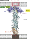
Cartoon of Vps13 bound between membranes and interacting with Mcp1p via its VAB. The chorein-N (orange), extended-chorein (teal), and APT1 (yellow) segments are believed to form a lipid transport channel. The VAB interacts with the Pro-X-Pro motif in the predicted unstructured N-terminus of Mcp1p (indicated). We speculate that the APT1 segment interfaces directly with the integral membrane domain of Mcp1, which harbors scrambling activity, and that there might be a direct handoff of lipids between Vps13p and the scramblase. The ribbons cartoon of the VPS13 lipid transport module was generated using RoseTTAFold (Baek et al., 2021).

References

    1. Ashkenazy, H., Abadi S., Martz E., Chay O., Mayrose I., Pupko T., and Ben-Tal N.. 2016. ConSurf 2016: An improved methodology to estimate and visualize evolutionary conservation in macromolecules. Nucleic Acids Res. 44:W344–W350. 10.1093/nar/gkw408 - DOI - PMC - PubMed
    1. Baek, M., DiMaio F., Anishchenko I., Dauparas J., Ovchinnikov S., Lee G.R., Wang J., Cong Q., Kinch L.N., Schaeffer R.D., et al. . 2021. Accurate prediction of protein structures and interactions using a three-track neural network. Science. 373:871–876. 10.1126/science.abj8754 - DOI - PMC - PubMed
    1. Bean, B.D.M., Dziurdzik S.K., Kolehmainen K.L., Fowler C.M.S., Kwong W.K., Grad L.I., Davey M., Schluter C., and Conibear E.. 2018. Competitive organelle-specific adaptors recruit Vps13 to membrane contact sites. J. Cell Biol. 217:3593–3607. 10.1083/jcb.201804111 - DOI - PMC - PubMed
    1. Dziurdzik, S.K., Bean B.D.M., Davey M., and Conibear E.. 2020. A VPS13D spastic ataxia mutation disrupts the conserved adaptor-binding site in yeast Vps13. Hum. Mol. Genet. 29:635–648. 10.1093/hmg/ddz318 - DOI - PMC - PubMed
    1. Dziurdzik, S.K., and Conibear E.. 2021. The Vps13 family of lipid transporters and its role at membrane contact sites. Int. J. Mol. Sci. 22:2905. 10.3390/ijms22062905 - DOI - PMC - PubMed

Publication types

MeSH terms