. 2022 Apr 22;50(7):3867-3891.
doi: 10.1093/nar/gkac213.

hnRNP G/RBMX enhances HPV16 E2 mRNA splicing through a novel splicing enhancer and inhibits production of spliced E7 oncogene mRNAs

Affiliations

hnRNP G/RBMX enhances HPV16 E2 mRNA splicing through a novel splicing enhancer and inhibits production of spliced E7 oncogene mRNAs

Chengyu Hao et al. Nucleic Acids Res. .

Abstract

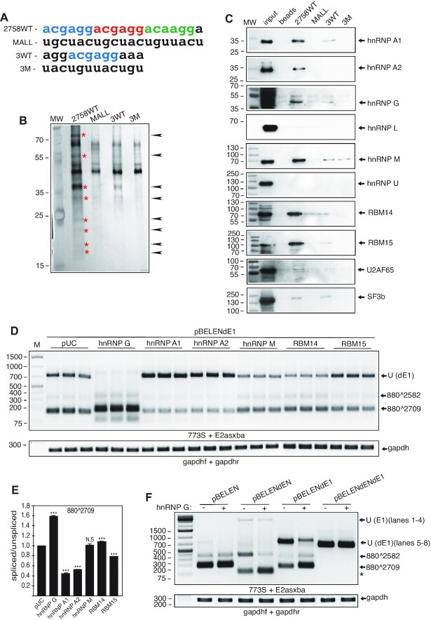
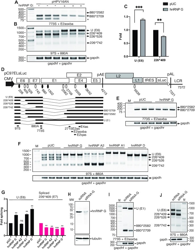
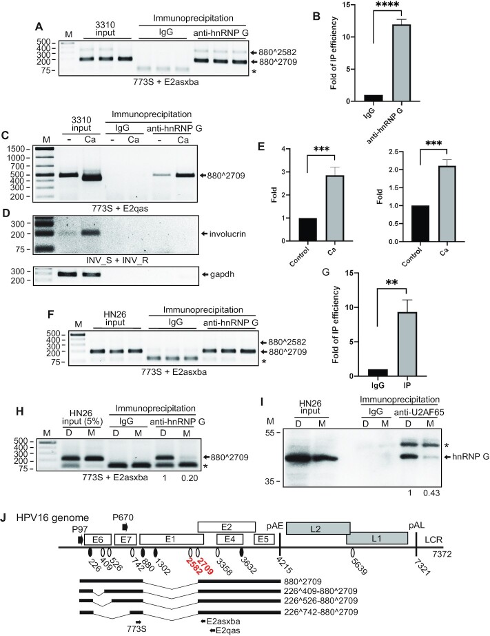
Human papillomavirus type 16 (HPV16) E2 is an essential HPV16 protein. We have investigated how HPV16 E2 expression is regulated and have identifed a splicing enhancer that is required for production of HPV16 E2 mRNAs. This uridine-less splicing enhancer sequence (ACGAGGACGAGGACAAGGA) contains 84% adenosine and guanosine and 16% cytosine and consists of three 'AC(A/G)AGG'-repeats. Mutational inactivation of the splicing enhancer reduced splicing to E2-mRNA specific splice site SA2709 and resulted in increased levels of unspliced E1-encoding mRNAs. The splicing enhancer sequence interacted with cellular RNA binding protein hnRNP G that promoted splicing to SA2709 and enhanced E2 mRNA production. The splicing-enhancing function of hnRNP G mapped to amino acids 236-286 of hnRNP G that were also shown to interact with splicing factor U2AF65. The interactions between hnRNP G and HPV16 E2 mRNAs and U2AF65 increased in response to keratinocyte differentiation as well as by the induction of the DNA damage response (DDR). The DDR reduced sumoylation of hnRNP G and pharmacological inhibition of sumoylation enhanced HPV16 E2 mRNA splicing and interactions between hnRNP G and E2 mRNAs and U2AF65. Intriguingly, hnRNP G also promoted intron retention of the HPV16 E6 coding region thereby inhibiting production of spliced E7 oncogene mRNAs.


Figures

Graphical Abstract
Graphical Abstract
hnRNP G/RBMX inhibits splicing in the E6 coding region thereby reducing production of the spliced E7 mRNA, while enhancing E2 mRNA splicing through a splicing enhancer at the E2 splice site.
Figure 1.
Figure 1.
(A) Linearized HPV16 genome (numbers refer to the HPV16 reference strain GeneBank: K02718.1). Early and late genes are indicated. P97: HPV16 early promoter. P670: HPV16 late promoter. Black oval: 5′-splice site/splice donor. White oval: 3′-splice site/splice acceptor. Splice acceptors SA2582 and SA2709 located upstream of the E2 open reading frame are indicated in red. pAE: HPV16 early polyadenylation site. pAL: HPV16 late polyadenylation site. LCR: long control region. A subset of alternatively spliced HPV16 E2 mRNAs all utilizing HPV16 3′-splice site SA2709. (B) Middle panel: Schematic representation of the HPV16 subgenomic reporter plasmid pBEL. Transcription of the HPV16 sequences in the pBEL plasmid is driven by the human cytomegalovirus immediate early promoter (CMV). Early and late genes are indicated. P97: HPV16 early promoter. P670: HPV16 late promoter. Black oval: 5′-splice site/splice donor. White oval: 3′-splice site/splice acceptor. pAE: HPV16 early polyadenylation site. pAL: HPV16 late polyadenylation site. Numbers refer to the HPV16 reference strain (GeneBank: K02718.1). Upper panel: Blow up of the region downstream of HPV16 3′splice site SA2709. The deletion introduced between positions 2731 and 2922 in pBEL to produce pD200 is shown, the introduction of a unique XbaI restriction site at position 2731 is indicated and the insertion of antisense sequences between 2731 and 2922 is indicated with an arrow (pBELAS). (C) The major HPV16 mRNAs produced from pBEL are spliced from HPV16 5′-splice site SD880 to either 3′-splice site SA2582, SA2709 or SA3358 and polyadenylated at pAE. (D) RT-PCR with indicated primers on RNA extracted from HeLa cells transfected with pBEL or pD200. RT-PCR was performed in the absence (−) or presence (+) of RT as indicated. Unspliced E1 encoding mRNAs U (E1) mRNAs and mRNAs spliced between SD880 and SA2582, SA2709 or SA3358 are indicated to the right. Note that the two gapdh samples represent controls for pBEL and pD200 performed in the presence of RT. M, molecular weight marker. (E) PCR on 2-fold serially diluted cDNA obtained from RNA extracted from HeLa cells transfected with pD200 or pBEL. For pD200, cDNA was undiluted, 2-, 4-, 8- and 16-fold diluted. For pBEL, cDNA was 32-, 64-, 128- and 256- and 512-fold diluted. (F) RT-qPCR with primers 773S and 2715E2as was performed as described in Materials and Methods to quantitate relative E2 mRNA levels produced by pBEL and pD200 upon transfection of HeLa cells. P < 0.0001. (G) RT-PCR with the indicated primers on RNA extracted from HeLa cells transfected with pBELAS, pBELCM, pD200 or pBEL.
Figure 2.
Figure 2.
(A) Schematic representation of genomic HPV16 plasmid pHPV16AN. LoxP sites and HPV16 early (P97) and late (P670) promoters and early (pAE) and late (pAL) poly(A) signals are indicated. Positions of PCR primers 16S and 16A are indicated. The effect of the cre recombinase on pHPV16AN in transfected cells is illustrated. (B) RT-PCR with indicated primers on RNA extracted from HeLa cells transfected with pHPV16AN or pHPV16ANdEN in the presence of cre-expressing plasmid pCAGGS-nlscre. Transfections were performed in triplicates. M, molecular weight marker. (C) RT-qPCR with primers 773S and 2715E2as was performed as described in Materials and Methods to quantitate relative E2 mRNA levels produced by pHPV16AN or pHPV16ANdEN upon transfection of HeLa cells. P < 0.0001. (D) RT-PCR with indicated primers on RNA extracted from HeLa cells transfected with pHPV16AN or pHPV16ANdEN. Transfections were performed in triplicates. (E) Schematic representation of the HPV16 subgenomic pC97ELsLuc reporter plasmid. Transcription of the HPV16 sequences in the pC97ELsLuc plasmid is driven by the human cytomegalovirus immediate early promoter (CMV). HPV16 splice sites are indicated. Numbers refer to the HPV16 reference strain (GeneBank: K02718.1). Early and late polyadenylation signals pAE and pAL are indicated. A subset of HPV16 alternatively spliced, early mRNAs are indicated. Arrows represent RT-PCR primers. IRES, internal ribosome entry site; sLuc, secreted luciferase. The deletion of the splicing enhancer downstream of splice site SA2709 is indicated at the top. (F–H) RT-PCR on RNA extracted from HeLa cells transfected with the indicated plasmids. RT-PCR primers are indicated below each gel image and spliced mRNAs represented by the amplicons are indicated to the right. (I) Densitometric quantification of electrophoresed RT-PCR products were performed as described in Materials and Methods. Percentage spliced E2 mRNA over unspliced/intron retained E1 mRNAs is shown. (J) RT-qPCR with primers 773S and 2715E2as was performed as described in Materials and Methods to quantitate relative E2 mRNA levels produced by pC97ELsLuc and pC97ELdEN upon transfection of HeLa cells. P < 0.0001.
Figure 3.
Figure 3.
(A) Sequences of biotinylated HPV16 RNA oligonucleotides used in RNA-mediated protein pull-downs of proteins from HeLa cell nuclear extracts. The three AC(G/A)AGG - repeats that constitute the enhancer are colored in the wild type RNA sequence (2758WT). MALL, all nucleotide positions in the wild type enhancer are mutated; 3WT, contains one AC(G/A)AGG – repeat; 3M, contains one mutant enhancer motif. Names of RNA oligonucleotides are listed to the left. (B) Silver stained SDS-polyacrylamide gel displaying proteins pulled down by the biotinylated RNA oligos shown in (A). Bands indicated with red stars and arrows were cut out from the gel and subjected to mass spectrometry. MW, molecular weight marker. (C) Western blotting of factors pulled down by Streptavidin-coated magnetic beads carrying biotinylated HPV16 RNA oligo 2758WT, MALL, 3WT or 3M. Filters were stained with antibodies to proteins identified by mass spectrometry (indicated to the right) and by antibodies to hnRNP L and hnRNP U that were not identified by mass spectrometry and served as negative controls. (D) RT-PCR with primers 773S and E2asxba on RNA extracted from HeLa cells transfected with HPV16 subgenomic plasmid pBELENdE1 in the presence of empty pUC plasmid or plasmids expressing either of the indicated proteins. Unspliced U (dE1) mRNA and mRNAs spliced between SD880 and SA2582 or SA2709 are indicated to the right of the gel image. Transfections were performed in triplicates. Locations of RT-PCR primers in HPV16 reporter plasmid pBELENdE1 are shown in Supplementary Figure S3C. M, molecular weight marker. (E) Densitometric quantification of RT-PCR bands in (A). The quantitations are displayed as ratios between cDNA bands representing HPV16 mRNAs spliced between SD880 and SA2709 and cDNA bands representing unspliced HPV16 mRNAs (U (dE1)) in each transfected sample. Splicing ratio monitored with pBELENdE1 cotransfected with pUC was set as 1. Quantitations were performed on triplicate samples. Statistically significant differences between 880∧2709 cDNA levels in samples transfected with pBELENdE1 and plasmids expressing either of the indicated proteins and samples transfected with pBELENdE1 and empty pUC plasmid are shown. ***, P < 0.001; N.S., not significantly different. (F) RT-PCR with primers 773S and E2asxba on RNA extracted from HeLa cells transfected with HPV16 subgenomic plasmids pBELEN, pBELENdEN, pBELENdE1 or pBELENdENdE1 in the absence (−) or presence (+) of hnRNP G plasmid. Unspliced U (dE1) mRNA and mRNAs spliced between SD880 and SA2582 or SA2709 are indicated to the right of the gel image. *, position of primer-dimer products.
Figure 4.
Figure 4.
(A, B) RT-PCR with indicated primers on RNA extracted from HeLa cells transfected with pHPV16AN and cre-expressing plasmid in the absence (−) or presence (+) of phnRNP G. Triplicate transfections are shown. (A) mRNAs spliced between SD880 and SA2582 or SA2709 are indicated to the right of the gel image. *, position of primer-dimer products. (B) Unspliced/intron retained U (E6) mRNA and mRNAs spliced between SD880 and SA409, SA526 or SA742 are indicated to the right of the gel image. (C) Densitometric quantification of RT-PCR bands (U (E6) and 226∧409) in (B) in the absence or presence of hnRNP G. The The quantitations are displayed as relative levels of U (E6) cDNAs or 226∧409 in the absence or presence of hnRNP G. (D) Schematic representation of the HPV16 subgenomic pC97ELsLuc reporter plasmid. A subset of HPV16 alternatively spliced, early mRNAs are indicated. Arrows represent RT-PCR primers. IRES, internal ribosome entry site; sLuc, secreted luciferase. (E) RT-PCR with indicated primers on RNA extracted from HeLa cells co-transfected with pC97ELsLuc and empty pUC plasmid or hnRNP G-expression plasmid. mRNAs spliced between SD880 and SA2582 or SA2709 are indicated to the right of the gel image. (F) RT-PCR with primers 97S and 880A on RNA extracted from HeLa cells co-transfected with pC97ELsLuc and empty pUC plasmid or hnRNP-expressing plasmids hnRNP G, hnRNP A2, hnRNP A1 or hnRNP D. Unspliced U (E6) mRNA and mRNAs spliced between SD880 and SA409, SA526 or SA742 are indicated to the right of the gel image. (G) Densitometric quantification of RT-PCR bands (U (E6) and 226∧409) in (H) in the absence or presence of hnRNP G. The quantitations are displayed as relative levels of U (E6) cDNAs or 226∧409 cDNAs in the absence or presence of hnRNP G. p-values are indicated. (H) Western blotting of hnRNP G in HeLa cells transfected with scrambled siRNAs (scRNA) or siRNAs to hnRNP G (siRNA-G). (I, J) HeLa cells were transfected with HPV16 subgenomic expression plasmid pC97ELsLuc and scrambled siRNA (scRNA) or siRNAs to hnRNP G, (siRNA-G) and RNA was extracted. (I) RT-PCR was performed with primers 773S and E2asxba. Unspliced/intron retained U (E1) mRNAs and mRNAs spliced between SD880 and SA2582 or SA2709 are indicated to the right. (J) RT-PCR was performed with primers 97S and 880A. Unspliced U (E6) mRNA and mRNAs spliced between SD880 and SA409, SA526 or SA742 are indicated to the right of the gel image.
Figure 5.
Figure 5.
(A) Schematic representation of the 5′-end of the HPV16 genome encoding E6, E7 and part of E1. HPV16 splice sites SD226, SA409, SA526, SA742 and SD880 are indicated. A previously identified splicing silencer is indicated as a red box with ‘S’. (B) Schematic representation of the HPV16 subgenomic expression plasmid pX856F in which HPV16 sequences encoding E6, E7 and part of E1 have been inserted downstream of the CMV promoter. HPV16 splice sites SD226, SA409, SA526, SA742 and SD880 are indicated. Regions and endpoints of sequences included in various HPV16 subgenomic expression plasmids are indicated. Locations of the RT-PCR primers 97S and X478A are indicated. (C) RT-PCR with primers 97S and X478A on RNA extracted from HeLa cells co-transfected with the indicated HPV16 plasmids and empty pUC plasmid or hnRNP G-expressing plasmid. Unspliced/intron retained U (E6) mRNA and mRNAs spliced between SD880 and SA409 are indicated to the right of the gel image. (D) Densitometric quantitation of triplicate samples in the gel images in (C). Quantitations are displayed as %-splicing in the graph. Mean values and standard deviations are shown. ns, not statistically significant difference. (E) Western blotting on cell extracts from HeLa cells co-transfected with pC97ElsLuc and pUC or hnRNP G plasmid. Cells were harvested 20hrs posttransfection. Blots were stained with antibody specific for hnRNP G or tubulin as described in Materials and Methods. (F) Densitometric quantification of the hnRNP G band in (E) in the absence or presence of transfected hnRNP G plasmid. The quantitations are displayed as relative levels of hnRNP G in the absence or presence of transfected hnRNP G plasmid. P < 0.01. (G) Western blotting on cell extracts from HeLa cells transfected with HPV16 reporter plasmid pHAE6E7Flag and empty pUC plasmid or hnRNP G expression plasmid. Blots were stained with antibody specific for HA-tag to detect HA-tagged HPV16 E6 protein or tubulin. (−), untransfected cells. (H) Western blotting on cell extracts from HeLa cells transfected with HPV16 reporter plasmid pHAE6E7Flag and empty pUC plasmid or hnRNP G expression plasmid. Blots were stained with antibody specific for FLAG-tag to detect FLAG-tagged HPV16 E7 protein or tubulin. Results from triplicate transfections are shown. (I) Densitometric quantification of the FLAG-E7 band in (H) in the absence or presence of transfected hnRNP G plasmid. The quantitations are displayed as relative levels of FLAG-E7 in the absence or presence of transfected hnRNP G plasmid. P < 0.01.
Figure 6.
Figure 6.
(A) Schematic representation of plasmids encoding wild type hnRNP G or various hnRNP G mutants. Amino acids positions in the hnRNP G proteins are indicated. RRM, RNA recognition motif; RGG, arginine-glycine-glycine motif-rich sequence (RGG-box); NTD, nascent RNA targeting domain; serine-arginine-glycine-tyrosine-motif rich domain (SRGY-box); C-RBD, C-terminal RNA binding domain. (B) Western blotting on cell extracts from HeLa cells transfected with empty pUC plasmid or plasmids expressing either of the various Flag-tagged hnRNP G proteins displayed in A. Blots were stained with antibody specific for Flag-tag to detect Flag-tagged hnRNP G proteins or with tubulin-specific antibody. (C) RT-PCR with primers 773S and E2asxba on RNA extracted from HeLa cells co-transfected with pBELENdE1 and empty pUC plasmid or plasmids expressing wild type or mutant hnRNP G. Unspliced/intron retained U (dE1) mRNAs and mRNAs spliced between SD880 and SA2582 or SA2709 are indicated to the right. Locations of RT-PCR primers in HPV16 reporter plasmid pBELENdE1 are shown in Supplementary Figure S3C. (D) RT-PCR with primers 97S and 880as on RNA extracted from HeLa cells cotransfected with pC97ELsLuc and empty pUC plasmid or plasmids expressing wild type or mutant hnRNP G. Unspliced/intron retained U (E6) mRNA and mRNAs spliced between SD880 and SA409, SA526 or SA742 are indicated to the right of the gel image. Locations of RT-PCR primers in HPV16 reporter plasmid pC97ELsLuc are shown in Figure 4D. (E) Densitometric quantification of RT-PCR bands (U (dE1) or 880∧2709) in (C). The The quantitations are displayed as relative levels of U (dE1) cDNAs or 880∧2709 in the absence or presence of hnRNP G. Transfections with empty pUC plasmid is set as 1. P-values are indicated. (F) Densitometric quantification of RT-PCR bands (U (E6), 226∧409 or 226∧742) in (D). The The quantitations are displayed as relative levels of U (E6) cDNAs, 226∧409 or 226∧742 in the absence or presence of hnRNP G. Transfections with empty pUC plasmid is set as 1. P-values are indicated.
Figure 7.
Figure 7.
(A) RT-PCR with primers 773S and E2asxba on RNA extracted from HeLa cells co-transfected with pBELENdE1 and empty pUC plasmid or plasmids expressing wild type or mutant hnRNP G. Unspliced/intron retained U (dE1) mRNAs and mRNAs spliced between SD880 and SA2582 or SA2709 are indicated to the right. Locations of RT-PCR primers in HPV16 reporter plasmid pBELENdE1 are shown in Supplementary Figure S3C. (B) Densitometric quantification of RT-PCR bands (U (dE1) or 880∧2709) in (C). The The quantitations are displayed as relative levels of U (dE1) cDNAs or 880∧2709 in the absence or presence of hnRNP G. Transfections with empty pUC plasmid is set as 1. P-values are indicated. (C) RT-PCR with primers 97S and 880as on RNA extracted from HeLa cells cotransfected with pC97ELsLuc and empty pUC plasmid or plasmids expressing wild type or mutant hnRNP G. Unspliced/intron retained U (E6) mRNA and mRNAs spliced between SD880 and SA409, SA526 or SA742 are indicated to the right of the gel image. Locations of RT-PCR primers in HPV16 reporter plasmid pC97ELsLuc are shown in Figure 4D. (D) Densitometric quantification of RT-PCR bands (U (E6), 226∧409 or 226∧742) in (D). The quantitations are displayed as relative levels of U (E6) cDNAs, 226∧409 or 226∧742 in the absence or presence of hnRNP G. Transfections with empty pUC plasmid is set as 1. P-values are indicated.
Figure 8.
Figure 8.
(A) Immunoprecipitation of proteins in cell extracts from untransfected (−) HeLa cells or from HeLa cells cotransfected with plasmids encoding U2AF65 (+) and hnRNP G (+) using either IgG or anti-hnRNP G antibody followed by Western blotting with antibodies to either U2AF65 or hnRNP A1. Input samples were subjected to Western blotting with antibodies to U2AF65, hnRNP A1, hnRNP G or tubulin. M, molecular weight marker. (B) Western blotting on cell extracts of HeLa cells transfected with plasmid encoding wild type, flag-tagged hnRNP G or the indicated flag-tagged hnRNP G mutants. Western blotting was performed with anti-Flag antibody, anti-U2AF65 antibody or anti-tubulin antibody. (C) Coimmunoprecipitation experiment on cell extracts from HeLa cells transfected with pUC-plasmid or Flag-tagged hnRNP G or the indicated Flag-tagged hnRNP G mutants using anti-U2AF65 antibody followed by Western blotting with anti-Flag antibody. (D) RT-PCR with primers 773S and E2qas on RNA extracted from C33A2 cells treated with the indicated concentrations of melphalan for 22 h. Locations of RT-PCR primers in HPV16 reporter plasmid pBELsLuc in the C33A2 cells are shown in Supplementary Figure S7B and C. (E) Densitometric quantitation of the RT-PCR bands in (D) that represent HPV16 unspliced/retained intron U (E1) mRNAs or mRNAs spliced from SD880 to SA2582 or to SA2709. (F) RT-PCR with primers 773S and E2qas on RNA extracted from C33A2 cells treated with the indicated concentrations of melphalan for the indicated time periods. cDNAs representing HPV16 mRNAs spliced between SD880 and SA2582 or SA2709 are indicated to the right. (G) Western blotting on cell extracts from C33A2 cells treated with DMSO or 50uM melphalan for 20hrs in triplicates. Blots were stained with antibody specific for hnRNP G or tubulin. (H) Densitometric quantitation of the Western blots in D. The quantitations are displayed as relative levels of hnRNP G levels in melphalan-treated cells over DMSO-treated cells. (I) C33A2 cells were treated with DMSO or melphalan for 21 h, lysed in RIPA buffer and subjected to immunoprecipitation with IgG or anti-hnRNP G antibody followed by extraction of the immunoprecipitated RNA and RT-PCR with HPV16-specific primers 773s and E2asxba. Input represents RT-PCR with the same primers on RNA extracted from 5% of the input in the immunoprecipitation incubation. cDNAs representing HPV16 mRNAs spliced between SD880 and SA2582 or SA2709 are indicated to the right. *, primer dimers. (J) Densitometric quantitation of the RT-PCR bands in (I) that represent HPV16 E2 mRNAs spliced from SD880 to SA2709. The quantitations are displayed as relative levels of 880∧2709 cDNAs obtained from anti-hnRNP G immunoprecipitated RNA-protein complexes obtained from melphalan-treated cells over DMSO-treated cells. (K) Immunoprecipitation of proteins in cell extracts from DMSO (D) or melphalan (M)-treated C33A2 cells using either IgG or anti-U2AF65 antibody followed by Western blotting with antibody to hnRNP G. M, molecular weight marker. (L) HeLa cells transfected with HPV16 reporter plasmid pBEL in the absence or presence of hnRNP G plasmid were lysed in RIPA buffer and subjected to immunoprecipitation with IgG or anti-U2AF65 antibody followed by extraction of the immunoprecipitated RNA and RT-PCR by HPV16 specific primers 773s and E2asxba. Input represents RT-PCR with the same primers on RNA extracted from 5% of the input in the immunoprecipitation incubation. M, molecular size marker; *, primer dimers. (M) RT-qPCR with primers 773S and E2asxba was performed as described in Materials and Methods on the RNA analyzed in (L). **, P < 0.001.
Figure 9.
Figure 9.
(A) Immunoprecipitation of proteins in cell extracts from DMSO (D) or melphalan (M) treated C33A2 cells using either IgG or anti-P(S/T/Y) antibody followed by western blotting with antibody to hnRNP G. Input samples represent 5% of the sample volume used for immunoprecipitation. MW, molecular weight marker; *, unknown band. (B) Immunoprecipitation of proteins in cell extracts from DMSO (D) or melphalan (M) treated C33A2 cells using either IgG or anti-sumo antibody followed by western blotting with antibody to hnRNP G. (C) C33A2 cells were treated with DMSO (D) or 10- or 100uM of sumoylation inhibitor 2-D08 followed by RNA extraction and RT-PCR with HPV16-specific primers 773s and E2qas. Locations of RT-PCR primers in HPV16 reporter plasmid pBELsLuc in the C33A2 cells are shown in Supplementary Figure S7B and C. (D) Densitometric quantification of the RT-PCR band in (C) representing HPV16 880∧2709 spliced mRNAs. The quantitations are displayed as relative levels of 880∧2709 cDNAs in 2-D08-treated cells over DMSO-treated cells. P-values are indicated. (E) C33A2 cells were treated with DMSO (D) or 10- or 100 uM of sumoylation inhibitor 2-D08 followed by lysis in RIPA buffer and immunoprecipitation with IgG or anti-hnRNP G antibody. RNA was extracted from the Immunoprecipitated RNA-protein complexes and subjected to RT-PCR with HPV16-specific primers 773s and E2asxba. Locations of RT-PCR primers in HPV16 reporter plasmid pBELsLuc in the C33A2 cells are shown in Supplementary Figure S7B and C. *, primer dimer. (F) Immunoprecipitation of proteins in cell extracts from DMSO (D) or sumoylation inhibitor 2-D08-treated C33A2 cells using either IgG or anti-U2AF65 antibody followed by western blotting with antibody to hnRNP G. Input samples represent 10% of the sample volume used for immunoprecipitation.
Figure 10.
Figure 10.
(A) HPV16-immortalized human keratinocyte cell line 3310 cells were lysed in RIPA buffer and subjected to immunoprecipitation with IgG or anti-hnRNP G antibody followed by extraction of the immunoprecipitated RNA and RT-PCR by HPV16 specific primers 773s and E2asxba. Input represents RT-PCR with the same primers on RNA extracted from 5% of the input in the immunoprecipitation incubation. M, molecular size marker; *, primer dimers. (B) Densitometric quantitation of the RT-PCR bands in (A) that represent HPV16 E2 mRNAs spliced from SD880 to SA2709. The quantitations are displayed as relative levels of 880∧2709 cDNAs obtained from anti-hnRNP G immunoprecipitated RNA–protein complexes over IgG immunoprecipitated complexes. (C) HPV16-immortalized human keratinocyte cell line 3310 cells were treated with 2.4 mM CaCl2 to induce differentiation, lysed in RIPA buffer and subjected to immunoprecipitation with IgG or anti-hnRNP G antibody followed by extraction of the immunoprecipitated RNA and RT-PCR by HPV16 specific primers 773s and E2asxba. Input represents RT-PCR with the same primers on RNA extracted from 5% of the input in the immunoprecipitation incubation. (D) RT-PCR with primers specific for mRNAs encoding the cell differentiation marker involucrin performed on the cDNA samples used in (C). (E) Densitometric quantitation of the RT-PCR bands in (C) that represent HPV16 E2 mRNAs spliced from SD880 to SA2709. The quantitations are displayed as relative levels of 880∧2709 cDNAs obtained from CaCl2-treated cells over untreated cells. (F) Cells from the HPV16-positive tonsillar cancer cell line HN26 were lysed in RIPA buffer and subjected to immunoprecipitation with IgG or anti-hnRNP G antibody followed by extraction of the immunoprecipitated RNA and RT-PCR by HPV16 specific primers 773s and E2asxba. Input represents RT-PCR with the same primers on RNA extracted from 5% of the input in the immunoprecipitation incubation. M, molecular size marker; *, primer dimers. (G) Densitometric quantitation of the RT-PCR bands in (F) that represent HPV16 E2 mRNAs spliced from SD880 to SA2709. The quantitations are displayed as relative levels of 880∧2709 cDNAs obtained from anti-hnRNP G immunoprecipitated RNA-protein complexes over IgG immunoprecipitated complexes. (H) HN26 cells were treated with DMSO (D) or melphalan (M) for 21 h, lysed in RIPA buffer and subjected to immunoprecipitation with IgG or anti-hnRNP G antibody followed by extraction of the immunoprecipitated RNA and RT-PCR with HPV16-specific primers 773s and E2asxba. Input represents RT-PCR with the same primers on RNA extracted from 5% of the input in the immunoprecipitation incubation. cDNAs representing HPV16 mRNAs spliced between SD880 and SA2709 are indicated to the right. *, primer dimers. (I) Immunoprecipitation of proteins in cell extracts from DMSO (D) or melphalan (M) treated HN26 cells using either IgG or anti-U2AF65 antibody followed by western blotting with antibody to hnRNP G. Input samples represent 10% of the sample volume used for immunoprecipitation. (J) Linearized HPV16 genome (numbers refer to the HPV16 reference strain GeneBank: K02718.1). Early and late genes are indicated. P97: HPV16 early promoter. P670: HPV16 late promoter. Black oval: 5′-splice site/splice donor. White oval: 3′-splice site/splice acceptor. Splice acceptors SA2582 and SA2709 located upstream of the E2 open reading frame are indicated in red. pAE: HPV16 early polyadenylation site. pAL: HPV16 late polyadenylation site. LCR: long control region. A subset of alternatively spliced HPV16 E2 mRNAs all utilizing HPV16 3′-splice site SA2709. Arrows indicate RT-PCR primers used to detect the HPV16 E2 mRNAs in HPV16 immortalized 3310 cells or tonsillar cancer cell line HN26.
Figure 11.
Figure 11.
Schematic representation of the HPV16 genome. The early P97 promoter and the late P670 promoter are indicated. HPV16 splice sites and early and late polyadenylation signals pAE and pAL, respectively, are indicated. hnRNP G interacts with HPV16 sequences in the E6/E7 coding region that are present on HPV16 mRNAs produced from the HPV16 early promoter P97 and promotes retention of the E6-coding intron, at the same time inhibiting splicing and inhibiting production of the E7 mRNAs and E7 protein. On HPV16 mRNAs lacking E6/E7 sequences, i.e. mRNAs produced from the HPV16 late promoter P670, hnRNP G enhances splicing to E2 mRNA-specific 3′-splice site SA2709 which results in increased E2 mRNA production. Increased E2 protein levels would contribute to a transcriptional shut-down of the early HPV16 P97 promoter further reducing production of E7 mRNAs and E7 protein thereby paving the way for cell differentiation and induction of late gene expression.

Similar articles

Cited by

References

    1. Walboomers J.M., Jacobs M.V., Manos M.M., Bosch F.X., Kummer J.A., Shah K.V., Snijders P.J., Peto J., Meijer C.J., Munoz N.. Human papillomavirus is a necessary cause of invasive cervical cancer worldwide. J. Pathol. 1999; 189:12–19. - PubMed
    1. Bouvard V., Baan R., Straif K., Grosse Y., Secretan B., El Ghissassi F., Benbrahim-Tallaa L., Guha N., Freeman C., Galichet L.et al. .. A review of human carcinogens–Part B: biological agents. Lancet Oncol. 2009; 10:321–322. - PubMed
    1. Kajitani N., Satsuka A., Kawate A., Sakai H.. Productive lifecycle of human papillomaviruses that depends upon squamous epithelial differentiation. Front. Microbiol. 2012; 3:152. - PMC - PubMed
    1. Chow L.T., Broker T.R., Steinberg B.M.. The natural history of human papillomavirus infections of the mucosal epithelia. APMIS. 2010; 118:422–449. - PubMed
    1. Schiffman M., Doorbar J., Wentzensen N., de Sanjose S., Fakhry C., Monk B.J., Stanley M.A., Franceschi S.. Carcinogenic human papillomavirus infection. Nat. Rev. Dis. Primers. 2016; 2:16086. - PubMed

MeSH terms

Substances